
Introduction
The inception of percutaneous coronary intervention (PCI) dates back to 1977 when Andreas Grüntzig performed the first coronary balloon angioplasty, revolutionizing the treatment of coronary artery disease.1 Since then, PCI has undergone significant advances, including the development of drug-eluting stents (DES),2 which parallel enhancements in intravascular imaging technologies. One of the earliest and most pivotal technologies is intravascular ultrasound (IVUS), which Yock and colleagues pioneered in the 1980s.3
Since then, IVUS has become a crucial tool in the arsenal of contemporary interventional cardiology, and IVUS-guided PCI has significantly reduced major adverse cardiovascular events (MACE), including mortality, target lesion failure (TLF), myocardial infarction (MI), and stent thrombosis.4,5 These benefits are consistent across different risk profiles, lesion types, and generations of stents.4 Consequently, the European Society of Cardiology (ESC) and the American Heart Association/American College of Cardiology (ACC/AHA) have upgraded intracoronary imaging guidance by IVUS or optical coherence tomography to class I for performing PCI on anatomically complex lesions, particularly left main disease, true bifurcations, and long lesions.6,7
However, despite robust data supporting its effectiveness and endorsements from clinical guidelines, the adoption of IVUS in everyday practice exhibits notable variation, influenced by regional practices, the individual experience of operators, and economic factors. Enhancing its usage in clinical settings hinges on improved education regarding its applications, clearer guideline recommendations, and better reimbursement practices. This review offers an in-depth perspective on the evidence and clinical utility of IVUS, emphasizing its transformative potential in refining the quality of interventional cardiology practices.
Fundamentals of the IVUS
IVUS employs principles similar to general ultrasound imaging, where piezoelectric crystals produce ultrasound pulses activated by electrical current.8 These pulses are reflected by tissue structures within the vessel walls, and the echoes are captured to create grayscale images. Highly reflective or echogenic structures like fibrous tissues and calcifications appear as brighter areas, while less reflective, echo-lucent structures such as lipid pools appear as darker images.9
The resolution of IVUS depends on several factors, including the wavelength and the beam width, because shorter wavelengths and narrower beam widths enhance axial and lateral resolutions. The typical axial resolution for these systems is around 80 μm to 100 μm, with lateral resolution of approximately 200 μm to 250 μm and tissue penetration between 6 mm and 12 mm.9,10 Advanced high-definition IVUS systems are now available, offering transducer frequencies up to 60 MHz, which provide even superior axial resolution, less than 40 μm—important for imaging modern thin-strut stents, which typically have strut thicknesses of 60 μm to 80 μm.8
There are two primary types of IVUS catheters used in contemporary clinical settings. The first is the mechanical rotational single transducer probe, typically operating at 40 MHz to 45 MHz sound frequency, such as the OptiCross™ by Boston Scientific. The second type is the phased-array probe, which features multiple fixed transducers and operates at a lower frequency of about 20 MHz, like the Eagle Eye™ from Philips Volcano. Both types of catheters can be used through 5F or 6F guiding catheters, with sizes ranging from 2.6F to 3.5F. Automatic or manual image pullbacks are commonly performed at a standard speed of 1.0 or 0.5 mm/s. Table 1 describes the characteristics of different IVUS catheters.
Table 1
Characteristics of intravascular ultrasound catheters.
| CHARACTERISTICS | BOSTON SCIENTIFIC OPTICROSS | PHILLIPS VOLCANO EAGLE EYE | PHILLIPS VOLCANO REVOLUTION | PHILLIPS VOLCANO REFINITY | ACIST KODAMA | TERUMO VIEWIT |
|---|---|---|---|---|---|---|
| Transducer frequency | 40 MHz (50 MHz HD also available) | 20 MHz | 45 MHz | 45 MHz | 60 MHz | 40 MHz |
| Distal shaft profile | 3.1F | 3.3F | 3.2F | 3F | 3.2F | 2.6F |
| Proximal shaft profile | 3.1F | 2.9F | 3.5F | 3F | 3.6F | 3.2F |
| Transducer to tip length | 20 mm | 10 mm (2.5 mm for short-tip version) | 30 mm | 20.5 mm | 20 mm | 29 mm |
| Guiding catheter | ≥ 5F | ≥ 5F | ≥ 6F | ≥ 5F | ≥ 6F | ≥ 5F |
Evidence Supporting IVUS-Guided PCI
Several randomized controlled trials and meta-analyses (Table 2) have demonstrated the benefits of intravascular imaging-guided PCI. The IVUS-XPL (Impact of Intravascular Ultrasound Guidance on the Outcomes of Xience Prime Stents in Long Lesions) trial, which included 1,400 participants with long lesions, demonstrated a reduction in MACE over 5 years with IVUS.11 However, in the ULTIMATE (Intravascular Ultrasound Guided Drug Eluting Stents Implantation in “All-Comers” Coronary Lesions) study, IVUS-guided PCI yielded a significant reduction in MACE in patients with a wide variety of lesions.12
Table 2
Characteristics of recent meta-analyses of randomized controlled trials. IVUS: intravascular ultrasound; OCT: optical coherence tomography; MI: myocardial infarction; TLR: target-lesion revascularization
| AUTHOR/ YEAR | NO. OF TRIALS AND PATIENTS | OUTCOMES |
|---|---|---|
| Khan at al./ 20234 | 20 trials and 11,698 patients comparing intravascular imaging guided PCI vs angiography guided PCI | Cardiac death, MI, stent thrombosis, target vessel revascularization, and TLR |
| Kuno et al./ 202344 | 32 trials and 22,684 patients comparing intravascular imaging guided, functionally guided vs angiography guided PCI | Trial defined MACE (composite of cardiovascular death, MI, and TLR) |
| Giacoppo et al./ 202413 | 24 trials (15,489 patients: IVUS vs angiography, 46.4%; OCT vs angiography: 32.1%; IVUS vs OCT: 21.4%) | The two co-primary outcomes were TLR and MI |
| Stone et al./202431 | 22 trials and 15,694 patients comparing IVUS vs OCT vs angiography-guided PCI | TLR, defined as the composite of cardiac death, target vessel MI, or TLR |
A recent meta-analysis of randomized controlled trials demonstrated that intravascular imaging-guided PCI was associated with a 47% relative risk reduction in cardiac death, 19% in MI, 56% in-stent thrombosis, 26% in target vessel revascularization, and 29% decrease in target lesion revascularization.4 These benefits corresponded to the lesion complexity and patient risk, with high-certainty evidence showing that intravascular imaging could result in 23 to 64 fewer cardiac deaths, 15 to 19 fewer MIs, 9 to 13 fewer stent thromboses, 28 to 38 fewer target vessel revascularizations, and 35 to 48 fewer target lesion revascularizations per 1,000 people treated, depending on the patient’s risk category.3
Other network meta-analyses have demonstrated similar observations.5,13 An analysis of Medicare beneficiaries from 2009 to 2017 showed that while IVUS was used in only 10.5% of all PCI procedures, its use gradually increased, particularly in academic centers and for complex lesions.14 This trend correlated with improved outcomes, including lower mortality risks, MI, and repeat revascularization compared to angiography-guided PCI. However, despite these benefits, the proportion of IVUS usage in the United States (US) remains considerably lower than in countries like Japan, where it was reported to be around 84% (Figure 1).15
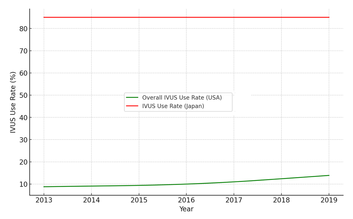
Figure 1
Trends of intravascular ultrasound (IVUS) use in the United States and Japan. To compare the IVUS use rates in percutaneous coronary intervention between the USA and Japan from 2013 to 2019, we extracted data points from provided graphical charts in prior publications.14 For Japan, the data covered up to 2017, which we extrapolated to 2019 using linear extrapolation based on the last observed rate.15 Both datasets were interpolated for smooth transitions between years.
Role in Complex Lesions
In complex coronary interventions (ie, left main disease, diffuse disease, chronic total occlusion (CTO), in-stent restenosis, ostial and bifurcation lesions) and low-contrast PCI, the clinical benefits of IVUS are notably prominent. The MAIN-COMPARE (Revascularization for Unprotected Left Main Coronary Artery Stenosis: Comparison of Percutaneous Coronary Angioplasty versus Surgical Revascularization) registry highlighted the efficacy of IVUS-guided stenting, which was utilized in more than 77% of PCI cases, demonstrating that IVUS-guided PCI for left main coronary artery disease was comparable to coronary artery bypass grafting in terms of all-cause mortality and a composite end point of death, MI, or stroke at 10 years.16 A detailed propensity score-matched analysis from this study revealed that IVUS-guided PCI resulted in a 60% reduction in all-cause mortality at 3 years compared with angiography-guided procedures.17 Another analysis from a Korean registry involving 487 matched pairs showed that IVUS-guided PCI significantly lowered the incidence of death or MI in non-left main bifurcation lesions compared to angiography-guided PCI.18
IVUS also plays a critical role in managing bifurcation PCI, particularly in determining the strategy for side branches. In these procedures, a preintervention minimal lumen area (MLA) of > 2.4 mm² at the side-branch ostium is a sensitive predictor of achieving a fractional flow reserve (FFR) of at least 0.80 after main branch crossover stenting.19
IVUS is indispensable in PCI for CTO, where it resolves proximal cap ambiguity, delineates cap morphology, and confirms the position of guidewires in relation to the true lumen. The CTO-IVUS (Chronic Total Occlusion InterVention with drUg-eluting Stents) study demonstrated a significant reduction in MACE with IVUS guidance compared to angiography-only guidance, emphasizing its value in complex PCI settings.20
IVUS has become an essential imaging modality for performing PCI in patients where contrast use must be minimized or eliminated, such as patients with severe renal impairment or contrast allergy.21 Coregistration is a key technique that facilitates low-contrast PCI, which integrates angiographic imaging with IVUS to provide real-time, high-resolution vessel assessment.22 In conventional PCI, IVUS-guided procedures rely on contrast-enhanced angiography for lesion localization, but a modified workflow is required in low-contrast settings. The coregistration process begins with selecting a reference frame from an index angiogram acquired at the end-diastolic phase, ensuring minimal cardiac motion artifacts.22 Automated software determines the ideal IVUS pullback trajectory within the target vessel. During IVUS pullback, ECG-triggered fluoroscopy is performed in synchronization with cardiac cycles, allowing the system to match the IVUS transducer’s location to the corresponding angiographic reference, thereby constructing an accurately mapped vessel image with minimal contrast injection.
In ultra-low contrast PCI, the coregistration technique is further adapted using an intracoronary wire-based angiographic view instead of a traditional contrast-enhanced angiogram.23 This approach enables precise lesion localization by leveraging the guidewire’s spatial reference, reducing or eliminating the need for contrast agents. The software estimates the IVUS catheter’s position relative to the inserted guiding catheter, creating a co-registered fluoroscopic-IVUS image that facilitates accurate stent sizing, optimal landing zone selection, and post-stent optimization.
Practical Implications
IVUS-guided PCI Optimization
Lesion Evaluation
Studies have demonstrated the role of hemodynamic assessment (FFR or instantaneous Wave Free Ratio [iFR]) in assessing the significance of coronary stenoses, revealing a weak correlation between angiographic and physiological assessments of stenosis severity.24, 25, 26 This has implications for IVUS, especially in evaluating MLA measurements, which show a modest correlation with FFR. Despite this, IVUS is vital in assessing left main disease where hemodynamic assessment limitations are pronounced. Research has identified optimal MLA thresholds that correlate with FFR values, recommending non-left main lesion cutoffs of around 4.0 mm² and left main artery cutoffs closer to 6.0 mm².27 In the FLAVOUR (FFR and Intravascular Ultrasound-Guided Intervention Strategy for Clinical Outcomes in Patients with Intermediate Stenosis) trial, FFR-guided PCI was non-inferior to IVUS-guided PCI among patients with intermediate stenosis.28
Plaque Morphology
IVUS also excels in detecting and characterizing plaque (Figure 2), surpassing angiography in identifying calcified lesions, which predict problems such as stent delivery failure and under-expansion. The technique quantifies calcium by measuring the angle and length of the calcified arc, termed the calcium index, which assists in determining the necessary mode and extent of plaque modification before stent implantation.27 IVUS also distinguishes between soft lipid-rich plaques, which are echolucent and predisposed to in-stent restenosis, and hard fibrous plaques, which show greater echogenicity. This differentiation is crucial because it influences the choice of treatment strategies, such as using cutting balloons for hard plaques and considering potential microembolization in lipid-rich plaques during PCI.
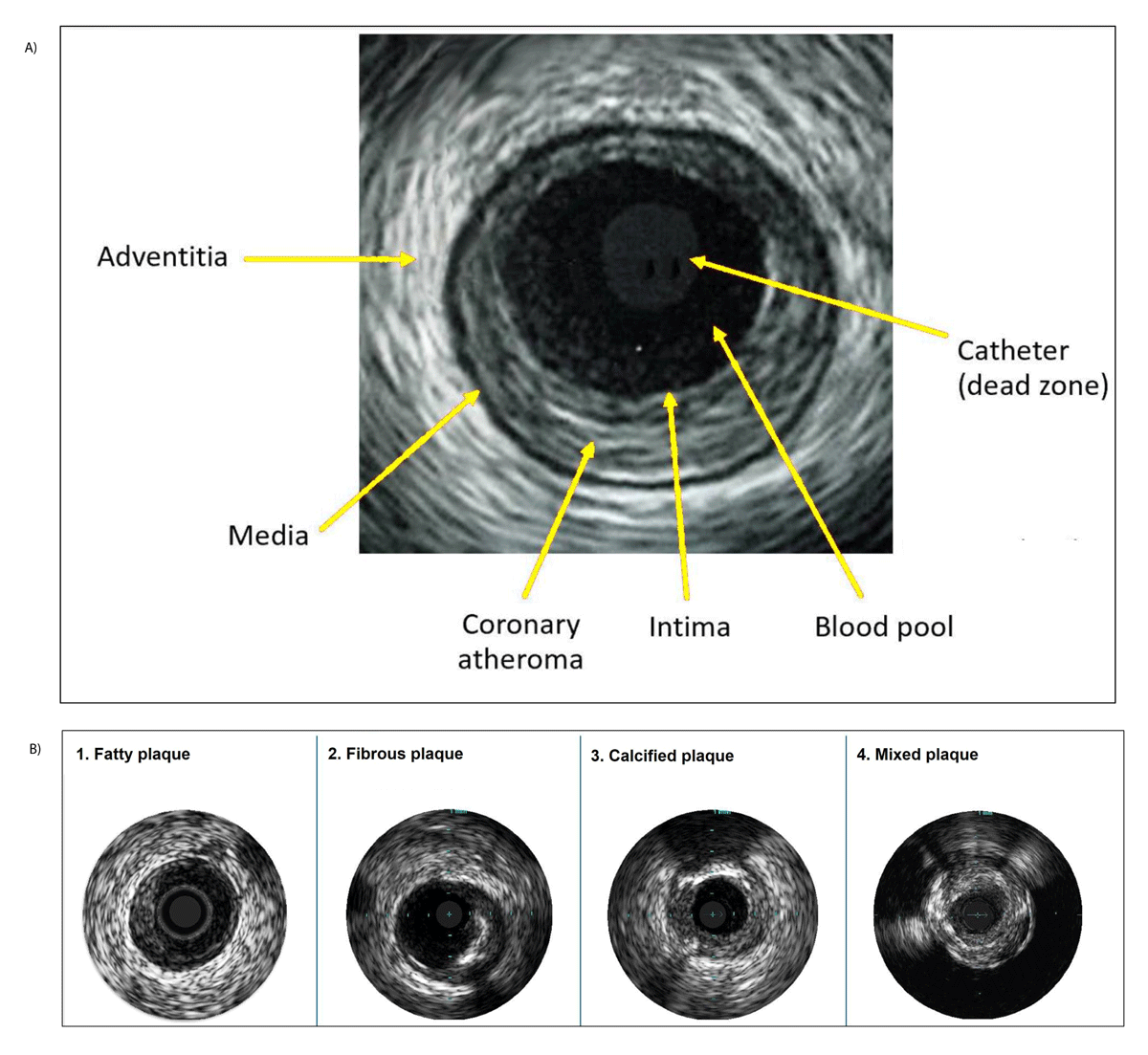
Figure 2
Intravascular ultrasound images of (A) vessel structure and (B) different plaque types.
Plaque Vulnerability
IVUS is instrumental in quantifying plaque burden, which is defined as the ratio of atheroma to the area encompassed by the external elastic.29 This measurement is instrumental in patients with positive or negative remodeling.30 Distinct from luminal stenosis, this metric is vital in assessing cardiovascular risk, with studies such as PROSPECT (The Providing Regional Observations to Study Predictors of Events in the Coronary Tree) linking a plaque burden of 70% or more in nonculprit lesions to increased MACE.31 Additionally, radiofrequency analysis with virtual histology IVUS offers insights into the presence of thin-cap fibroatheromas, with larger plaques significantly associated with higher MACE, underscoring the utility of IVUS in both research and clinical contexts for enhancing prognostic assessments and therapeutic planning.32 In the recent PREVENT (Preventive percutaneous coronary intervention versus optimal medical therapy alone for the treatment of vulnerable atherosclerotic coronary plaques) trial, preventive PCI in patients with non-flow–limiting vulnerable plaque as identified by intravascular imaging was shown to reduce MACE compared with medical therapy.33
Stent Placement Planning
IVUS has been instrumental in refining stent placement strategies. By offering a detailed understanding of plaque characteristics, IVUS assists in determining the best stent landing zones so that there is adequate coverage of the atherosclerotic segment of the vessel, choosing the correct stent sizes, and reducing risks associated with stent underexpansion and malapposition. The technology provides a superior perspective to traditional two-dimensional angiography, especially vital in strategically important areas like the left main bifurcation.
Residual plaque burden post-intervention has been recognized as a significant independent predictor of restenosis, particularly when more than 50% of plaque remains at the stent margins.34 IVUS has shown that even angiographically “normal” coronary arteries typically exhibit a plaque burden of around 50%.35 Consequently, a stepwise approach is advocated for determining optimal stent placement. Ideally, stents should land in an IVUS-identified segment with normal characteristics or, when that is not feasible, in areas where the plaque burden is less than 50%.
IVUS also excels in vessel sizing and detecting vessel remodeling, which is crucial for selecting appropriate stent sizes. It enables accurate measurement of the vessel dimensions, including positive or negative remodeling, even in larger vessels. Various sizing techniques are employed, ranging from conservative lumen-based sizing to more aggressive approaches using the external elastic lamina. Depending on the chosen strategy, stent size adjustments might include rounding up by 0 mm to 0.25 mm for a lumen-based approach or rounding down to the nearest 0.25 mm if based on the external elastic lamina. This precision significantly enhances the procedural success and long-term efficacy of stent implantation.
The following section discusses several approaches for stent planning, including our unique SEE IVUS technique for lesion assessment and stent sizing using Eagle Eye™ from Philips Volcano catheters.
Approaches for Stent Planning
Automated Pullback Technique
The automatic pullback technique in IVUS imaging uses a motorized retraction system to ensure a uniform, continuous acquisition of cross-sectional images along the coronary artery. In modern IVUS systems, the catheter is secured to a specialized sled that facilitates a consistent, automated retraction of the imaging core within the vessel.36 Traditional systems typically operate with automated pullback speeds ranging from 0.5 mm/s to 1.0, while newer models have been developed to achieve speeds up to 10 mm/s, further enhancing imaging efficiency and diagnostic precision.37
Co-registration Software-Based Technique
Co-registration software integrates IVUS images with angiographic data to produce a synchronized, real-time mapping of the coronary anatomy alongside the deployed stent.38 This fusion allows clinicians to precisely correlate stent positioning with anatomical landmarks, facilitating the detection of issues like edge dissections, malapposition, and incomplete expansion.37
SEE IVUS Technique
Figure 3 illustrates the SEE IVUS stepwise approach—often referred to as the “poor man’s coregistration”—designed to quickly and accurately obtain three critical data points: (1) the healthiest or near healthy distal landing zone, (2) the healthiest or near healthy proximal landing zone, (3) plaque morphology, and (4) lesion length. Automated algorithms often provide excessive, unnecessary details, while sled pullback can be time-consuming.
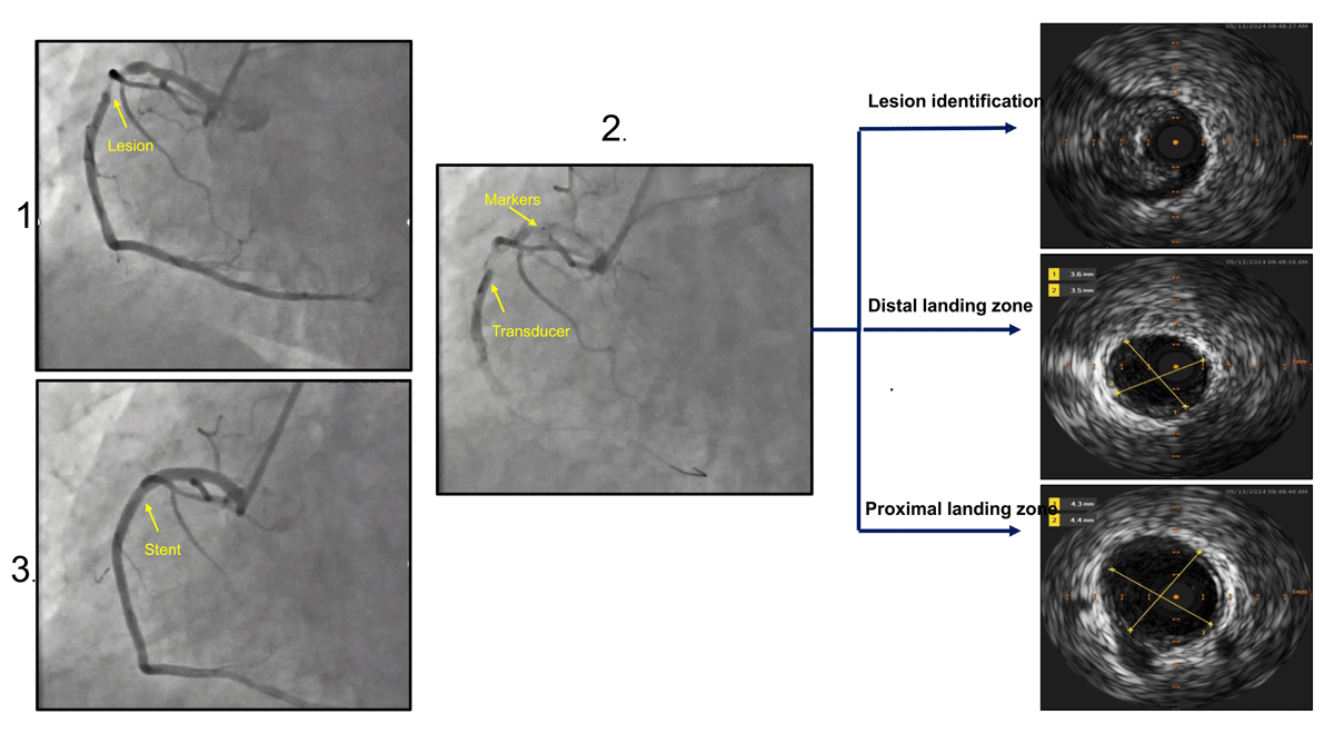
Figure 3
Stepwise approach of SEE IVUS technique. (1) Angiographic evaluation of lesion in the right coronary artery; (2) identification of stent length using IVUS after evaluating lesion, distal and proximal healthy landing zones; and (3) angiogram after stent placement. IVUS: intravascular ultrasound
The Philips Eagle Eye IVUS is advanced across the lesion to identify the optimal distal stent landing zone, targeting the healthiest or least diseased vessel. Contrast injection (5 cc for the right coronary artery and 10 cc for the left) with cine imaging is performed while noting side branches. IVUS catheter occlusion is not a concern. The proximal landing zone is determined angiographically, followed by an efficient pullback. Additional contrast cine can further refine the proximal stent landing zone if needed. If a distal stent is present, the IVUS tip is parked there, and the same steps are followed. If IVUS advancement is challenging, a buddy wire, guide extender technique, or further pretreatment should be considered.
The IVUS transducer is 5 mm radio-opaque and positioned 4 mm from the imaging element, with each marker representing 10 mm. The proximal and distal landing zones are confirmed with 2 cc to 4 cc contrast injection. Stent sizing is based on the distal healthy landing zone diameter and marker length to the proximal landing zone. The angiographic location of side branches is invaluable for precise placement.
This method offers key advantages: the stent behaves similarly to the IVUS catheter in tortuous vessels, ensuring accurate lesion coverage and reducing distal edge dissection. It also minimizes contrast, fluoroscopy, and cine use while enhancing stent delivery confidence—if the Eagle Eye crosses, a long stent almost always will. Additionally, it eliminates the need for complex algorithms or sled pullback; furthermore, the pullback speed can be fast or slow and does not affect the data gathered.
Post-PCI Evaluation
Post-PCI IVUS features have emerged as critical predictors of stent thrombosis, particularly stent underexpansion, persistent malapposition, significant edge dissections, and tissue prolapse (Figure 4).39,40 Among these, stent underexpansion, leading to a minimal stent area (MSA), has been most closely associated with early and late stent failure.41 The seminal MUSIC (Multicenter Ultrasound Stenting in Coronary Study) study, involving 155 stable patients, established a set of formal criteria for an optimal stent result after bare-metal stent implantation.41 Remarkably, these criteria were met in 81% of the cases, and even while administering only aspirin post-stenting, the stent thrombosis rate remained only 1.3% at 6 months.41
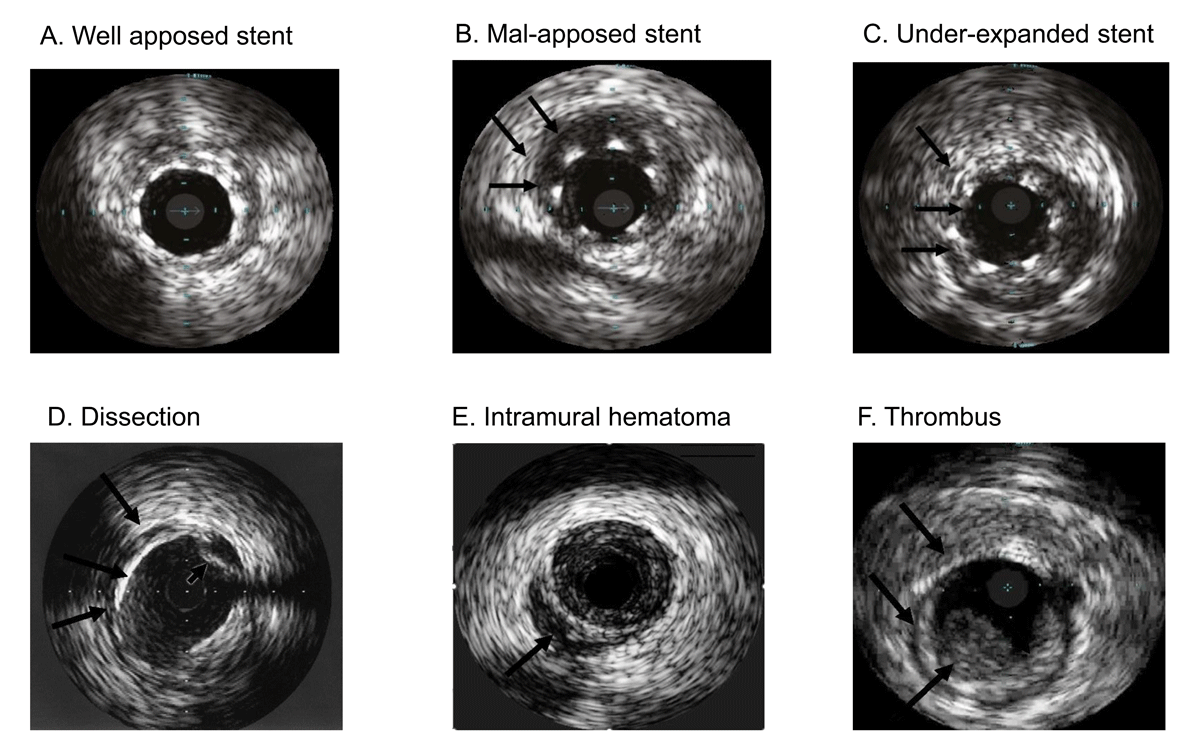
Figure 4
Intravascular ultrasound images of coronary stents and complications.
However, the MUSIC study criteria proved challenging to meet consistently, even in controlled trial settings. In the DES era, this challenge persisted despite the adoption of simpler IVUS criteria for optimal PCI outcomes. Large trials like IVUS-XPL (Impact of Intravascular Ultrasound Guidance on the Outcomes of Xience Prime Stents in Long Lesions) and ULTIMATE revealed that these simplified criteria were only met in about 50% of patients.12,42 Despite these challenges, the evidence consistently supports that even suboptimally achieved IVUS-guided DES PCI can lead to better outcomes than angiography-guided PCI, with more aggressive post-dilatation, larger MSAs, and improved clinical outcomes. Table 3 reports IVUS criteria used in different studies.
Table 3
Post stenting IVUS criteria used in landmark studies. MSA: minimal stent area; MLA: minimal lumen area
| STUDY | CRITERIA |
|---|---|
| MUSIC30 | 1. Complete strut apposition 2. Symmetrical stent expansion: minimum/maximum lumen diameter ≥ 0.7 3. Adequate stent expansion
|
| IVUS-XPL42 | 1. Minimal luminal cross-sectional area greater than the luminal cross-sectional area at the distal reference segment 2. Adequate stent expansion
|
| ULTIMATE12 | 1. An MLA in the stented segment > 5.0 mm2 or 90% of the MLA at the distal reference segments 2. Plaque burden 5 mm proximal or distal to the stent edge < 50%; and 3. No edge dissection involving the media with length more than 3 mm 4. Adequate stent expansion
|
| RENOVATE-COMPLEX PCI45 | Sufficient stent expansion without major stent malapposition to the vessel wall or edge dissection
|
There remains a clear need for a universal, evidence-based, and practical set of IVUS criteria that can be achieved across varied clinical settings. Earlier DES studies indicated that MSA less than 5.0 mm² to 5.5 mm² for non-left main lesions, less than 6 mm² for ostial LAD, less than 7 mm² for distal left main artery, and less than 8 mm² for left main trunk are predictive of fewer adverse events.27 Moreover, extensive dissections characterized by more than a 60° arc or more than 2 mm longitudinal extension, particularly those involving the media or adventitia, are associated with higher risks of stent thrombosis and should be avoided.9,43 Dissections or tissue protrusions resulting in an MLA of < 4 mm² are considered extensive and indicative of suboptimal stenting.43 While the clinical significance of acute stent malapposition remains uncertain, correcting any extensive acute stent malapposition exceeding 0.4 mm with a longitudinal extension over 1 mm wherever feasible is recommended.43
Conclusion
As PCI expands to include increasingly complex cases and diverse patient populations, the role of IVUS is poised to become even more integral. Advances such as radiofrequency tissue characterization, near-infrared spectroscopy, and iMap have moved beyond research into clinical relevance,8 with ongoing innovation expected to further improve the usability and integration of IVUS in daily practice. Moving forward, wider adoption of IVUS-guided strategies will be essential for optimizing outcomes, refining procedural planning, and driving progress in interventional cardiology.
Key Points
Intravascular ultrasound (IVUS) significantly reduces mortality and major adverse cardiovascular events by providing superior assessment of plaque morphology, vessel sizing, and stent placement, helping clinicians select optimal stent sizes, landing zones, and ensuring adequate expansion and apposition.
IVUS is especially valuable in managing complex scenarios such as left main disease, bifurcation lesions, chronic total occlusions, and procedures requiring minimal contrast, substantially improving procedural success and patient outcomes.
Consistent application of standardized IVUS optimization criteria—such as minimal stent area, plaque burden limits, and absence of significant dissections—is essential for achieving optimal outcomes and widespread adoption in clinical practice.
Competing Interests
Dr. Kleiman is a consultant for Abbott, Boston Scientific, Medtronic, and Siemens Health. The other authors have no competing interests to declare.