
Historical Overview
The first catheterization in humans was performed in 1929 by the German physician Werner Forssman, who performed right heart catheterization on himself using a urinary catheter.1 In 1963, the first transluminal angioplasty was done by Dr. Charles Dotter on a stenosed right iliac artery, setting the origins of catheter-based percutaneous treatment of occlusive arterial disease.2 In 1974, Dr. Andreas Grüntzig described “The Grüntzig balloon catheter principle” wherein a double-lumen catheter is used to apply uniform pressure for 10 to 30 seconds, compressing atherosclerotic plaque against the vessel wall and restoring blood flow. This became known as “percutaneous transluminal coronary angioplasty.” In 1977, he performed the first ever balloon dilatation of a coronary artery in humans.2 This has developed into one of the most successful therapeutic interventions in cardiovascular medicine.
Beyond ballooning, Dotter first described the concept of endovascular “splints” in 1964 and suggested using nonbiological endovascular stents to stabilize diseased arterial vessels.3 The first coronary stent was implanted in a patient by Dr. Jacques Puel in Toulouse, France, on March 28, 1986.4 The field of coronary artery stents also experienced enormous developments due to the efforts of Dr. Cesare Gianturco, Dr. Gary S. Roubin, Dr. Julio Palmaz, and Dr. Richard A. Schatz, all of whom made significant contributions to the development of balloon-expandable stents.5
Why Drug-Coated Balloons?
Percutaneous coronary intervention (PCI) using plain old balloon angiography (POBA), bare metal stents (BMS), or drug-eluting stents (DES) is the invasive treatment of choice for coronary artery disease. Limitations still remain, such as in-stent restenosis with BMS and restenosis with POBA.6 Consequently, a persistent concern—even with second-generation DES—has been the late occurrence of neoatherosclerosis and thrombotic events (6-8% for in-stent restenosis in the first year) and an adverse ischemic event rate of 2% per year with no apparent plateau.7 Furthermore, these adverse events are more frequently seen in certain situations, such as small-vessel disease and complex coronary bifurcation lesions with large stenosed side branches.8,9 Therefore, drug-coated balloons (DCB) emerged as an attractive alternative, fulfilling the “leaving nothing behind” strategy to eliminating the elevated thrombotic risk and increased bleeding risk from prolonged dual-antiplatelet therapy (DAPT) seen with permanent metallic prosthesis.
Drug-Coated Balloons
In the late 1990s, a research group in Tübingen, Germany, studied the effect of paclitaxel exposure on arterial walls, which demonstrated a dose-dependent inhibition of neointimal formation.10,11 In parallel, Drs. Ulrich Speck and Bruno Scheller demonstrated that the concomitant use of contrast agents served as carriers to paclitaxel and facilitated its transfer into the vessel wall.12,13,14 These experiences set the stage for the birth of DCB, where rapid-exchange semi-compliant balloon catheters coated with both an active antiproliferative substance—mainly paclitaxel and more recently sirolimus—and a carrier can permit rapid drug uptake and its persistence in vessel walls to inhibit neointimal formation.15 Figure 1 depicts the mechanism of DCB action in the vessel wall.
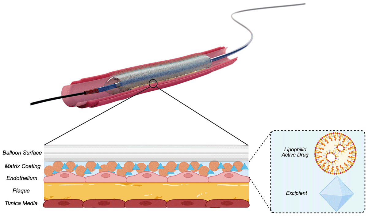
Figure 1
Drug-coated balloon mechanism of action. Reproduced with permission from Boston Scientific.
Several biological and procedural factors should be considered in effective DCB technologies. Antiproliferative agents can be divided into two main drugs: paclitaxel and sirolimus. Paclitaxel, a well-known potent antineoplastic agent, works by irreversibly binding to microtubules and enhancing microtubule assembly, which results in blockage of cell replication during mitosis. Consequently, reports have described paclitaxel as having a positive remodeling effect by late lumen increase post DCB-only therapy.16 Furthermore, paclitaxel is a highly lipophilic agent that enables rapid cellular uptake and work by maintaining a sustained biological effect at minimal doses.10
On the other hand, sirolimus, also known as rapamycin, possesses strong antiproliferative and immunosuppressant properties. It works by binding to intracellular cytosolic protein FK binding protein-12, forming a complex and reversibly inhibiting the activation of the mammalian Target of Rapamycin (mTOR), leading to inhibition of cell cycle progression at the G1/S transition.17 Certain limitations such as poor transfer rate and prolonged retention in the tissue are associated with sirolimus.18 Despite that, comparable clinical and angiographic treatment effects of paclitaxel coated balloon (PCB) and sirolimus coated balloon are described.19
Excipients, which are nonactive components, play a major role in active drug delivery and improving interaction with the vessel wall. Clinically proven carriers are iopromide, a low-osmolality contrast medium, and urea, an endogenous metabolite. These carriers improve drug adhesion and retention to warrant a prolonged therapeutic effect.
The development of a unique coating system is crucial to optimize drug delivery. The balloon coating properties offer advantages for drug release kinetics, adhesion properties, and overall efficacy. Coating features, such as having a homogenous thickness and drug concentration throughout a surface area, and coating fragmentation into submicron particles on balloon inflation are important to enhance the transfer capabilities of lipophilic drugs into the vascular tissue.20 Coating constituents significantly differ across devices, with different appearances for variations from resinous to multilayered and crystalline. A new approach with nano-encapsulative coatings may offer better drug solubility and stability with controlled release to enhance drug delivery. However, more robust clinical data is needed to progress this technology forward.
Due to differences in drug type, formulation, transfer kinetics, and bioavailability, DCBs are not considered to have a class effect, as variations in efficacy and safety profiles exist among different DCB options. Figure 2 highlights the main differences between paclitaxel and sirolimus DCB.
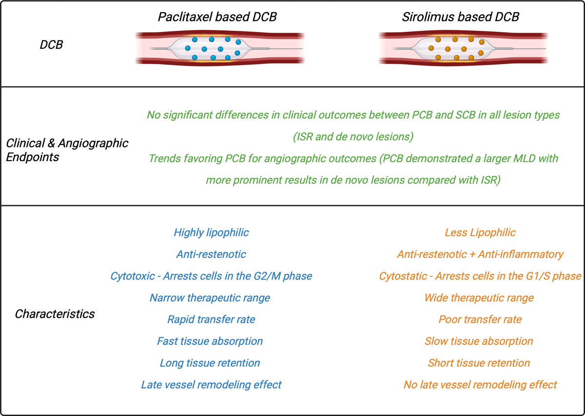
Figure 2
Main characteristics and differences between paclitaxel and sirolimus drug-coated balloons. DCB: drug-coated balloon; PCB: paclitaxel-coated balloon; SCB: sirolimus-coated balloon; ISR: in-stent restenosis; MLD: mean lumen diameter
In-Stent Restenosis
Following the introduction of BMS in the late 1990s, a new clinical entity appeared: in-stent restenosis (ISR). ISR is defined angiographically as > 50% reduction in the luminal diameter within a previously stented segment or adjacent to the stent edges (5 mm from the proximal and distal edges).21 Histologically, ISR is a slow process of negative vessel remodeling caused by exaggerated neointimal tissue changes. These changes are characterized as early ISR, mainly caused by neointimal hyperplasia and late ISR caused by neoatherosclerosis.22 Despite advancements in stent technology, which have led to a reduced risk ISR, it remains the leading cause of PCI failure, representing 5% to 10% of all PCI procedures in the US (United States).23 Therefore, it is essential to develop strategies to prevent and optimally manage ISR.
Table 1 highlights the main randomized controlled trials (RCTs) that showcase the performance of DCBs in ISR.24,25,26,27,28,29,30,31,32,33,34,35,36 The AGENT IDE (Clinical Trial to Assess the Agent Paclitaxel Coated PTCA Balloon Catheter for the Treatment of Subjects With In-Stent Restenosis) trial highlighted the superiority of DCB treatment strategy over POBA in mixed ISR. The study’s 1-year finding showed significantly reduced target lesion failure (TLF), target lesion revascularization (TLR), and target vessel-related myocardial infarction than in the POBA group, enabling Food and Drug Administration approval of the AGENT™ DCB (Boston Scientific Corporation) for ISR.25 Recently, the AGENT IDE trial’s 2-year data were also consistent with the prior findings, showing fewer events and repeat interventions with the DCB.37 Several other studies comparing DCB to POBA showed DCB’s superior angiographic and clinical outcomes.38,39 Figure 3 illustrates successful application of AGENT DCB for ISR.
Table 1
Summary of the main randomized clinical trials of the use of DCB in coronary ISR. BMS: bare metal stent; DCB: drug-coated balloon; DES: drug-coated stent; EES: everolimus-eluting stent; LLL: late lumen loss; MACE: major adverse clinical events; MLD: mean lumen diameter; ISR: in-stent restenosis; OCT: optical coherence tomography; PCB: paclitaxel-coated balloon; PES: paclitaxel-eluting stent; POBA: plain old balloon angioplasty; SCB: sirolimus-coated balloon; SES: sirolimus-eluting stent; TLF: target lesion failure; TVR: target vessel revascularization *P-value was not calculated for PES-PCB head-to-head comparison in late lumen loss.
| STUDY NAME | YEAR | ARMS | n | MAIN FINDINGS | MAIN CONCLUSION |
|---|---|---|---|---|---|
| BMS ISR | |||||
| PACOCATH ISR I & II24 | 2012 | POBA vs PCB | 108 | Angiography (6 months):
| Treatment of ISR with PCB is safe and consistently reduces repeat revascularization during long-term follow-up. |
| AGENT IDE25 | 2025 | POBA vs PCB | 600 | TLF (2 years): 34 % vs 27% (P = .04) | Consistent results were observed at 2-year follow-up, with a significantly reduced instance of TLF. |
| PEPCAD II26 | 2015 | PES vs PCB | 131 | Angiography (6 months):
| PCB consistently reduced LLL and restenosis compared to PES, indicating stability of the lesions treated. |
| RIBS V27 | 2016 | EES vs PCB | 189 | Angiography (6 months):
| EES was associated with superior angiographic effectiveness and reduces the need for target lesion revascularization compared to PCB. |
| SEDUCE28 | 2014 | EES vs PCB | 50 | Angiography (9 months):
| PCB was associated with better healing characteristics but appeared to be slightly less effective compared to EES. |
| TIS29 | 2016 | EES vs PCB | 136 | Angiography (12 months):
| Treatment of BMS ISR using PEB was associated with non-inferior angiographic results compared with the implantation of second-generation EES. |
| DES ISR | |||||
| PEPCAD-DES30 | 2012 | POBA vs PCB | 110 | Angiography (6 months):
| Angioplasty with PCB is superior to POBA in DES-ISR. |
| PEPCAD CHINA ISR31 | 2014 | PES vs PCB | 220 | Angiography (9 months):
| Angioplasty with DCB was noninferior to DES implantation in terms of angiographic effectiveness. |
| ISAR DESIRE 332 | 2013 | PES vs PCB | 268a | Angiography (6-8 months):
| Angioplasty with DCB was associated with noninferior effectiveness compared with DES implantation. |
| RIBS IV33 | 2015 | EES vs PCB | 309 | Angiography (6-9 months):
| In DES-ISR, EES provided superior long-term clinical and angiographic results compared with DEB. |
| RESTORE34 | 2018 | EES vs PCB | 172 | Angiography (9 months):
| In DES-ISR, both DCB and DES strategies were safe and effective up to 1-year post-procedure, with DES showing superior angiographic results. |
| Mixed ISR | |||||
| BIOLUX35 | 2018 | SES vs PCB | 229 | Angiography (6 months):
| In DES or BMS ISR, Angiographic results at 6 months and clinical outcomes up to 18 months post procedure are comparable between DCB and repeat DES groups. |
| DARE36 | 2018 | EES vs PCB | 278 | Angiography (6 months):
| In DES or BMS ISR, treatment with DCB is non-inferior to DES in terms of angiographic and clinical end points. |
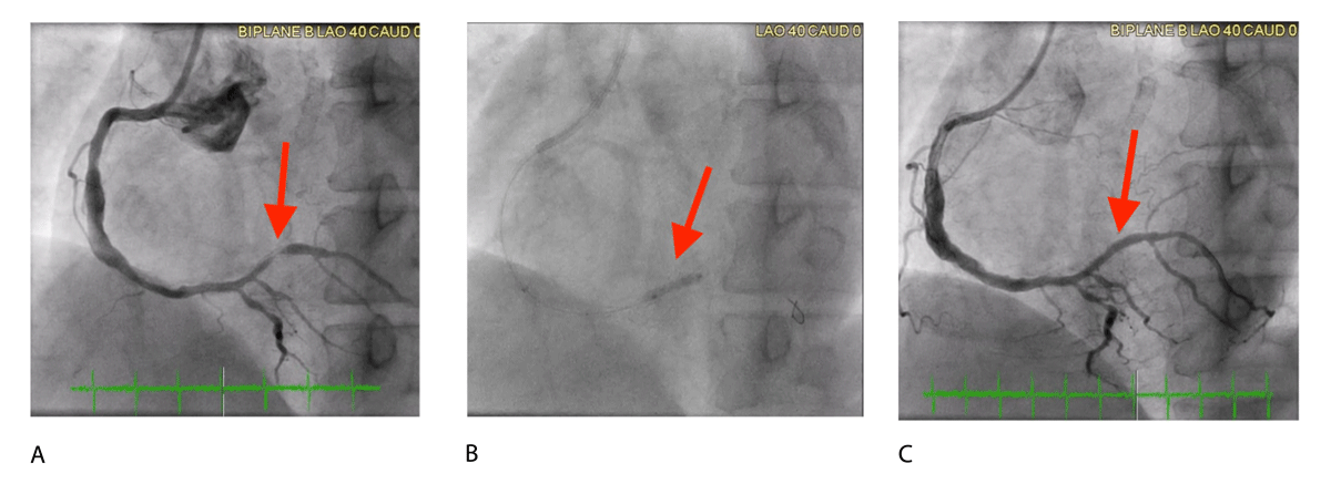
Figure 3
Coronary angiogram images demonstrating treatment of in-stent restenosis (ISR) using a drug-coated balloon (DCB). (A) Pre-treatment image showing 90% ISR in the affected artery. (B) Deployment of a 2.5 × 15 mm AGENT drug-coated balloon, inflated to 6 atm for 60 seconds. (C) Post-treatment image showing complete resolution of ISR with 0% stenosis after DCB intervention.
The DAEDALUS study (Difference in Anti-restenotic Effectiveness of Drug-eluting stent and drug-coated balloon Angioplasty for the occurrence of coronary in-Stent restenosis) was a large, investigator-initiated, collaborative, individual patient data meta-analysis that included 10 RCTs comparing DCB angioplasty with repeat stenting to DES DCBs for coronary ISR.40 At 3-year follow-up, repeat stenting with DES was moderately more effective than DCB with angioplasty at reducing the need for TLR. In addition, significant interaction was seen between treatment effect and type of restenosed stent, with more difference in patients with DES-ISR and comparable effects in patients with BMS-ISR. On the other hand, there was no significant difference in terms of the primary safety end point (composite of all-cause death, myocardial infarction, or target lesion thrombosis), with a numerically lower rate of this outcome in the DCB arm.
Subgroup analysis showed a significant correlation between treatment effect and generation of DES used to treat ISR, with a lower incidence of events associated with DCB compared with first-generation DES and similar effect between DCB and second-generation DES. In addition, a recent long-term nationwide analysis from the SCAAR (Swedish Coronary Angiography and Angioplasty Registry) showed similar findings to the DAEDALUS study, where DCB was associated with higher rates of TLR than DES. However, subgroup analysis between BMS-ISR and DES-ISR showed no significant difference between DES and DCB in terms of TLR and major adverse cardiac events (MACE).39 Therefore, DCB is equally effective and safe as repeat stenting with DES in treating BMS-ISR but less effective than repeat stenting with DES in treating DES-ISR. Based on that, the 2024 European Society of Cardiology/European Association for Cardio-Thoracic Surgery guidelines for the management of chronic coronary syndromes were updated and recommended repeat stenting with DES over DCB for the treatment of DES-ISR, with a Class I, Level A recommendation.41
Despite the findings of the DAEDALUS study, which showed inferiority of DCB compared to DES in DES-ISR, outcomes with DCB may vary based on differences in restenotic tissue and original stent characteristics.42 In real-world practice, specific anatomical and clinical scenarios may still favor a DCB approach. Situations such as small vessel size, stent underexpansion, complex bifurcations with side branches emerging from ISR segment, or multiple layers of previously implanted stent may limit the utility of an additional DES.
Although new-generation DES stents may slightly reduce the need for TLR in the ISR lesions,40 the risk of additional stent layers makes DCB the preferred choice for BMS- and DES-ISR when adequate lesion preparation is achieved post balloon angioplasty. Furthermore, patients who may benefit from a shorter duration of dual-antiplatelet therapy may be better candidates for DCB treatment. Therefore, patient-specific factors may influence outcomes and should guide treatment modality selection, as judged by the operator and targeted to the patient’s anatomy and clinical profile. Nevertheless, well-designed studies in different clinical and anatomic scenarios, powered for clinical end points and with long-term clinical follow-up, are warranted to refine the role of DCB in ISR.33,40
Studies on head-to-head comparisons of different DCBs showed no significant differences in angiographic and major clinical end points.43 Furthermore, some important measures may improve treatment efficacy of DCB in ISR, such as considering combined treatment of DCB with other devices,44,45 proper lesion preparation, and use of intravascular imaging and coronary physiology. Intravascular imaging with intravascular ultrasound or optical coherence tomography can provide insights into the mechanisms underlying ISR and currently have Class II, Level A recommendations.46,47 Identifying lesion-specific contributing mechanical factors can influence the decision between DCB and DES for the treatment of ISR. Consequently, intravascular imaging-guided lesion preparation before DCB use with aggressive pre-dilation with noncompliant balloons, cutting balloon, and high-pressure balloons can optimize DCB effect. Calcium modification tools such as rotablation, rotational atherectomy, lithotripsy, and scoring or cutting balloons may be adjunctive tools to improve stent expansion and luminal gain and to avoid slippage of pre-dilation balloons.44,48
Off-label Indications
Currently, DCBs are primarily indicated for the treatment of BMS and DES ISR. However, emerging evidence suggests expanding their use to include other indications, such as de novo coronary disease, bifurcation lesions, and diffuse long lesions. DCBs also may be considered for patients with diabetes, acute coronary syndrome, or those at high bleeding risk. Figures 4 and 5 include DCB indications and treatment strategy in coronary artery disease. The following sections explore the various off-label indications for DCB use, highlighting the growing body of evidence supporting its broader application.
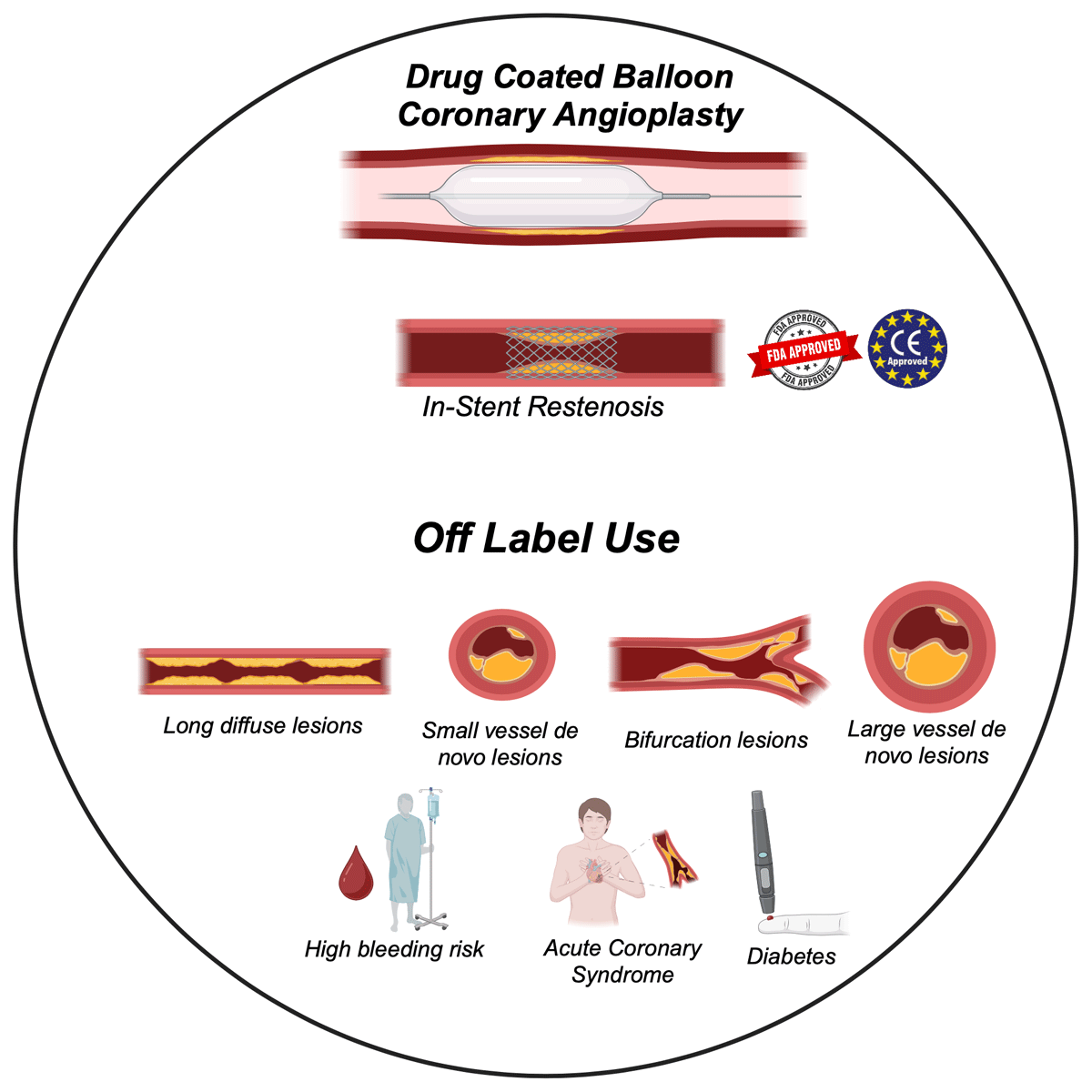
Figure 4
Drug-coated balloon coronary angioplasty indications.
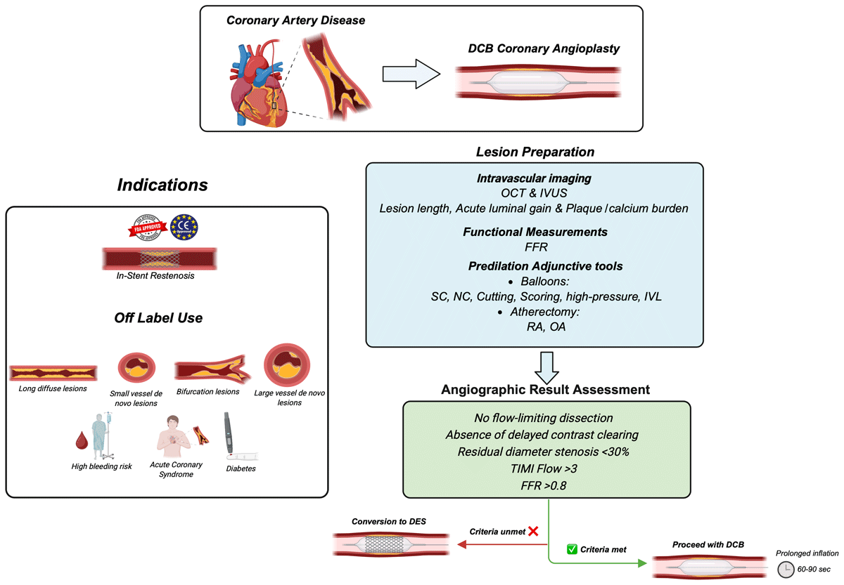
Figure 5
Central illustration: Drug-coated balloon indication and treatment strategy in coronary artery disease. Adopted from the international DCB consensus group. DCB: drug-coated balloon; DES: drug-eluting stent; FFR: fractional flow reserve; ISR: in-stent restenosis; IVL: Intravascular lithotripsy; IVUS: intravascular ultrasound; NC: non-compliant; OCT: optical coherence tomography; OA: orbital atherectomy; PCI: percutaneous coronary intervention; RA: rotational atherectomy; SC: semi compliant
De Novo Large Vessel Coronary Artery Disease
When treating large-caliber coronary artery disease vessels (LV-CAD), with diameters ≥ 3.0 mm, DESs have traditionally been the preferred option due to their ability to provide enhanced support to vessel walls. However, recent studies have shown that DCBs are also a viable treatment option. The DEBUT (Drug-Eluting Balloon in stable and Unstable angina: a randomized controlled non-inferiority) and PEPCAD-NSTEMI (Paclitaxel-Eluting PTCA-Balloon Catheter in Coronary Artery Disease for Treatment of Non–ST-Segment–Elevation Myocardial Infarction) trials both showed similar outcomes (TLF and MACE in both the DCB and metallic stent [BMS/DES] arms) among patients with high bleeding risk and acute coronary syndrome patients, respectively.49,50 It is important to note that the NSTEMI trial reported a 15% incidence of additional stent placement in the DCB group, highlighting the potential need for “bail-out” stenting due to unavoidable complications from DCBs, such as coronary dissection.50 Nevertheless, another retrospective study reported that while coronary dissection occurred in 28.3% of LV-CAD lesions, the need for bail-out stenting occurred in only 0.5% of cases, suggesting that most DCB-induced dissections are contained and can be medically managed.51
Additionally, use of DCB in LV-CAD has been studied in a variety of clinical contexts. The SPARTAN DCB trial and another retrospective study by Merinopoulos et al. showed no significant difference between primary DES and DCB in all-cause mortality when treating LV-CAD in the setting of stable CAD at 5- and 3.6-year follow-up, respectively.52,53 Merinopoulos et al. found similar outcomes—all-cause mortality, net cardiac events (TLF, cardiovascular mortality, subsequent acute coronary syndrome, CVA, TIA, and major bleeding)—among ST-segment elevation myocardial infarction (STEMI) patients between DCB-only and DES-only groups at 3 years.54 Furthermore, the REVELATION (Revascularization with Paclitaxel-Coated Balloon Angioplasty Versus Drug-Eluting Stenting in Acute Myocardial Infarction) trial evaluated the safety and efficacy of DCB angioplasty compared to DES in 120 patients presenting with STEMI and < 50% residual stenosis following successful pre-dilation. At 2-year follow-up, there was no significant difference in MACE between the two groups. Additionally, the DCB strategy was noninferior to DES in terms of fractional flow reserve (FFR) assessed at 9 months (0.92 ± 0.05 in the DCB group [n = 35] and 0.91 ± 0.06 in the DES group [n = 38; P = .27]).55 Although DCBs have gained interest as a monotherapy for LV-CAD, large randomized clinical trials that compare DCB to the gold standard of DES are needed to confirm their effectiveness and safety.
De Novo Small Vessel Coronary Artery Disease
Small vessel coronary artery disease is generally defined as a vessel diameter < 3.0 mm. Disadvantages arise when implanting stents in such a small vessel because late lumen loss over time becomes more detrimental, resulting in higher rates of ISR and adverse events.56 Early results from the single arm PEPCAD (Paclitaxel-Eluting PTCA-Balloon Catheter to Treat Small Vessel Coronary Artery Disease) study suggested favorability in safety and efficacy with low incidence of cardiac death, target vessel myocardial infarction, or TLR seen at 12 and 36 months.57
The PICCOLETO (Paclitaxel-coated balloon versus drug-eluting stent during PCI of small coronary vessels) trial showed contrasting results with higher percentage diameter stenosis compared to DES at 6-month follow-up.58
In the context of uncertainty surrounding optimal treatment strategies for small vessel coronary artery disease, the pivotal BELLO (Balloon Elution and Late Loss Optimization) trial provided important insights. This randomized study of 182 patients compared DCB angioplasty with paclitaxel-eluting stent (PES) implantation. At 6 months, DCB demonstrated both non-inferiority and statistical superiority over PES in reducing late lumen loss (0.08 ± 0.38 mm vs 0.29 ± 0.44 mm; P < .001 for non-inferiority, P = .001 for superiority), despite a significant bailout BMS implantation rate of 20%.59 At 24 months, the DCB group exhibited a significantly lower cumulative incidence of all-cause death, myocardial infarction, or target vessel revascularization compared to the PES group (14.4% vs 30.4%; P = .015). By 36 months, a significant reduction occurred in MACE with DCB (14.4%) versus PES (30.4%; P = .015),60 reinforcing the long-term clinical benefit of a stent-less strategy in selected patients with small-vessel disease.
The RESTORE SVD (Drug-Coated Balloon Versus Drug-Eluting Stent for Small-Vessel Disease) trial randomized 230 patients with de novo small vessel coronary disease to treatment with paclitaxel-coated balloon angioplasty or second-generation zotarolimus-eluting stents. At 9 months, the DCB group was noninferior to DES for the primary end point of in-segment diameter stenosis (29.6% ± 2.0% vs 24.1% ± 2.0%; P < .001). However, the DCB group had a smaller in-segment minimum lumen diameter (1.40 ± 0.42 mm vs 1.71 ± 0.39 mm; P < .001) and similar late lumen loss rates (0.25 ± 0.42 mm vs 0.27 ± 0.36 mm; P = .73) compared to the DES group.61 At 12 and 24 months, there were no significant differences in the composite clinical outcomes.
A large-scale observational analysis from the Swedish Coronary Angiography and Angioplasty Registry (SCAAR), which included 14,778 consecutive patients with de novo small-vessel coronary artery disease treated with either PCB or second-generation DES, reported contrasting findings. At three years, the risk of clinically significant restenosis was significantly higher in the PCB group compared to the DES group (adjusted HR, 2.03; 95% CI, 1.54-2.67). In contrast, the incidence of target lesion thrombosis did not differ significantly between the two groups (adjusted HR, 0.74; 95% CI, 0.41-1.33).62 Similarly, no significant differences were observed in all-cause mortality or myocardial infarction rates.
The discrepancy in the reported efficacy of DCBs for de novo small vessel CAD, along with inconsistent outcomes across multiple trials, may be attributed to the absence of a class effect among different DCB technologies and variations in procedural technique, particularly inadequate lesion preparation.57,58,59,61,63,64 For instance, the conflicting findings in the PICCOLETO (Drug Eluting Balloon Efficacy for Small Coronary Vessel Disease Treatment) study can be partially explained by suboptimal lesion pre-dilation, which was seen in approximately 75% of the patients enrolled.58 Table 2 provides a summary of the main RCTs of DCB in de novo small vessel coronary artery disease.58,59,64,65,66 Figures 6 and 7 illustrate our successful application of AGENT DCB for treatment of de novo small vessel CAD.
Table 2
Summary of the main clinical trials of the use of DCB in de novo small vessel coronary artery disease. PCB: paclitaxel-coated balloon; DES: drug coated stent; MACE: major adverse clinical events; DCB: drug coated balloon; LLL: late lumen loss; TLR: target lesion revascularization; BMS: bare metal stent
| STUDY NAME | YEAR | ARMS | n | MAIN FINDINGS | MAIN CONCLUSION |
|---|---|---|---|---|---|
| PICCOLETO58 | 2010 | PCB vs DES | 57 | Angiography (6 months):
| DCB angioplasty exhibited inferior antirestenotic efficacy compared to DES implantation. |
| BELLO59 | 2012 | PCB vs DES | 182 | Angiography (6 months)
MACE (6 months): 10% vs 16.3% (P = .021) | PCB shows lower angiographic late loss with comparable rates of restenosis and revascularization to DES. |
| BASKET-SMALL 265 | 2018 | PCB vs DES | 758 | Follow-up (12 months): Lesion-related MACE (12 months): 7.3% vs 7.5 % (P = .918) | PCB demonstrated non-inferiority to DES for MACE up to 12 months and similar event rates among both groups. |
| RESTORE SVD61 | 2018 | PCB vs DES | 230 | Angiography (9 months):
| PCB shows comparable clinical effectiveness and safety to DES implantation at 1 year. |
| PICCOLETO II64 | 2020 | PCB vs DES | 232 | Angiography (6 months):
| PCB showed noninferior angiographic effectiveness at 6 months and similar event rates at 1 year. |
| PEPCAD66 | 2013 | PCB vs BMS | 120 | Angiography (6 months)
MACE (12 & 36 months) 6.1% vs 37.5% (P < .001) | PCB demonstrated similar angiographic outcomes to BMS at 6 months with positive clinical outcomes of MACE and TLR at 1 and 3 years. |
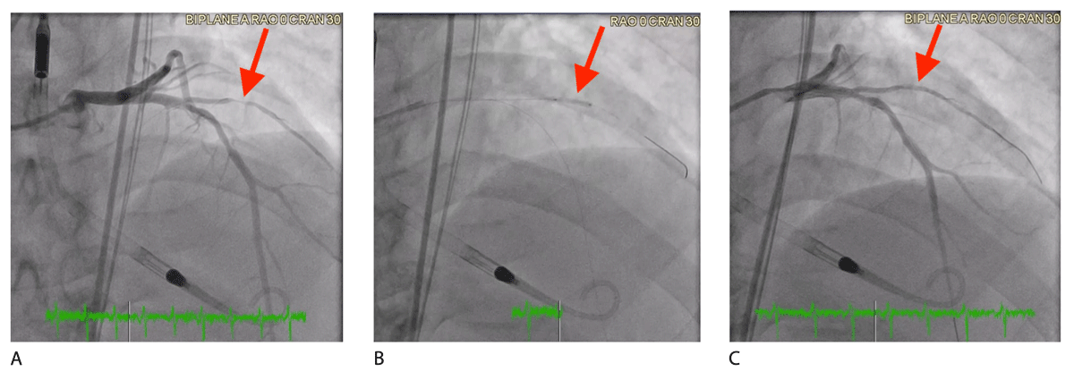
Figure 6
Coronary angiogram images illustrating treatment of de novo small vessel disease using a drug-coated balloon (DCB). (A) Pre-treatment image showing 90% stenosis proximal segment of the 1st diagonal branch. (B) Deployment of a 2.5 × 15 mm AGENT drug-coated balloon, inflated to 8 atm for 60 seconds. (C) Post-treatment image showing a significant reduction in stenosis to 20% following DCB intervention.
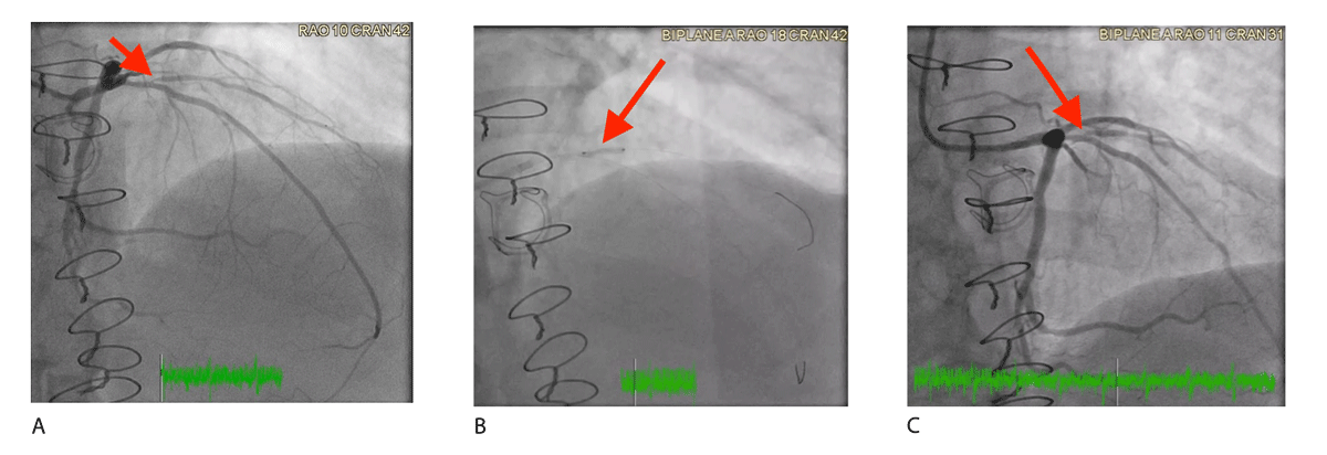
Figure 7
Coronary angiogram images demonstrating treatment of a de novo small vessel disease involving the ostial first diagonal branch with 90% stenosis using a drug-coated balloon (DCB). (A) Pre-treatment image showing 90% stenosis at the ostium of the first diagonal branch. (B) Deployment of a 2.5 × 12 mm AGENT DCB, inflated to 4 atm for 60 seconds. (C) Post-treatment image showing a reduction in stenosis to 30% and improved vessel patency following DCB intervention.
Bifurcation Lesions
Bifurcation lesions represent a significant challenge in coronary intervention, making up 15% to 20% of cases. These lesions are particularly difficult to manage due to the heightened risk of complications like branch vessel occlusion, restenosis, and stent thrombosis. Conventional double-stent placement for coronary bifurcation lesions offers no significant improvement in outcomes and may increase hospitalization and MACE rates.67 The most widely used approach remains single cross-over stenting in the main vessel, with side branch balloon dilation or provisional stenting performed as needed.
Single-stent placement remains the primary approach for treating bifurcation lesions, but it poses a risk of damaging side branches. POBA offers an alternative for expanding side branch vessels without modifying the main vessel structure. However, performing balloon-only PCI on side branch vessels after stenting the main branch still leads to a high restenosis rate in both branches.68 DCB preserves the benefits of POBA while offering an added potential for enhanced treatment efficacy, especially for small-size side branch vessels.
Several studies over the past decade have demonstrated promising results in treating bifurcation lesions using provisional stenting in the main branch and DCB on the side branch, as outlined in Table 3.69,70,71,72,73,74,75
Table 3
Summary of the main clinical trials of the use of DCB in coronary bifurcation lesions. BMS: bare metal stent; MB: main branch; DCB: drug coated balloon; SB: side branch; POBA: plain old balloon angioplasty; DES: drug coated stent; LLL: late lumen loss; MACE: major adverse clinical events; PCB: paclitaxel-coated balloon; TLR: target lesion revascularization
| STUDY NAME | YEAR | ARMS | n | MAIN FINDINGS | MAIN CONCLUSION |
|---|---|---|---|---|---|
| DEBIUT76 | 2012 | (A) BMS MB/DCB SB (B) BMS MB/POBA SB (C) DES MB/POBA SB | 117 | Angiography (6 & 12 months): LLL proximal MB, distal MB, distal SB (P = .001) LLL (A): 0.58 ± 0.65, 0.41 ± 0.60, and 0.19 ± 0.66 mm LLL (B): 0.60 ± 0.65, 0.49 ± 0.85, and 0.21 ± 0.57 mm LLL (C): 0.13 ± 0.45, 0.19 ± 0.64, and 0.11 ± 0.43 mm MACE (12 months)- A/B/C: 24.2%, 28.6%, 15% (P = .45) | Pretreatment of both MB and SB with PCB did not demonstrate any angiographic or clinical advantages over conventional BMS when using a provisional T-stenting approach. Additionally, DES showed better angiographic outcomes compared to both PCB and BMS. |
| PEPCAD-V70 | 2011 | BMS to MB & PCB to SB | 28 | Angiography (9 months): LLL: MB 0.38 ± 0.46, SB 0.21 ± 0.48 | Percutaneous treatment of bifurcation lesions with PCB to the SB was 100% successful and showed similar angiographic results to DES at 9 months. |
| PEPCAD-BIF71 | 2016 | PCB vs POBA of SB | 64 | Angiography (9 months): LLL: 0.13 mm 0.51 mm (P = .013) | PCB is a superior strategy to POBA of side branch vessels in bifurcation lesions. |
| BIOLUX-172 | 2015 | DES to MB & PCB to SB | 35 | Angiography (9 months): LLL of SB: 0.10 ± 0.43 | DES to the main branch and PCB to the side branch is a safe and effective strategy to treat bifurcation lesions. |
| BABILON75 | 2014 | (PCB) BMS MB & PCB SB vs (DES) DES MB & POBA SB* | 108 | Angiography (9 months): LLL (MB): PCB 0.31 ± 0.48 mm DES 0.16 ± 0.38 mm (P = .15) LLL (SB): PCB -0.04 ± 0.76 mm DES -0.03 ± 0.51 mm (P = .983) MACE (2 years): PCB 17.3% vs DES 7.1% (P = .045) | PCB bifurcation pretreatment with BMS in the MB resulted in higher LLL and more MACE events than everolimus DES, while both strategies had similar outcomes in the SB. |
| EASTBOURNE-BIF74 | 2024 | DES MB & PCB to SB vs de novo non-bifurcation lesions | 194 | Angiography (12 months): TLR: 4.2% vs 2% (P = .28) MACE 8.8% vs 5.2% (P = .081) | The use of this DCB, either alone or in combination with drug-eluting stents, could offer a safe and effective alternative to stents for treating bifurcations. |
[i] *NOTE: MB pre-dilation with PCB in the BMS MB group
The BABILON (Paclitaxel-Coated Balloon in Bifurcated Lesions) trial randomized 108 patients with coronary bifurcation lesions to either sequential pre-dilation with DCB angioplasty in both branches followed by provisional BMS implantation in the main branch, or to a standard approach with pre-dilation followed by provisional everolimus-eluting stent placement in the main branch. At 9 months, the DCB plus BMS strategy demonstrated noninferiority to the DES-alone strategy in terms of in-segment late lumen loss (0.31 ± 0.48 mm vs 0.16 ± 0.38 mm; P < .001 for noninferiority, P = .15 for superiority). Side branch late lumen loss was comparable between groups (–0.04 ± 0.76 mm vs –0.03 ± 0.51 mm; P = .983).75 However, the DCB group experienced a significantly higher rate of TLR at 9 months (13.5% vs 1.8%; P = .027), predominantly driven by restenosis in the main branch, and nonstatistical significance in terms of MACE.
Conflicting evidence was seen in the DEBIUT (Drug-Eluting Balloon in Bifurcations Trial) study, where the 6-month angiographic follow-up showed no significant differences in the DCB-based strategy compared to the BMS-based approach and was found to be less effective than the DES-based strategy.76
Another trial focused on the potential benefit of DCB angioplasty for the treatment of the SB after DES implantation in the main branch.71 Nine-month follow-up revealed significantly better results in the DCB group, where in-lesion late lumen loss was lower (0.13 ± 0.31 mm vs 0.51 ± 0.66 mm; P = .013), minimum lumen diameter was greater (1.78 ± 0.37 mm vs 1.39 ± 0.80 mm; P = .015), and binary restenosis was less frequent (5.9% vs 25.7%; P = .045) in patients treated with DCB.
As evidence supporting the use of DCB in bifurcation lesions continues to grow, its routine application for this indication may become more prevalent in daily practice. Nevertheless, more robust and high-quality evidence are still needed.
Diffuse Long Lesions
Stent length and burden (number of stents) are predictors of ISR and in-stent thrombosis. This directly applies to diffuse long lesions. This specific coronary phenotype necessitates the use of long stents or even overlapping stents, both of which are associated with an increased risk of compromising long-term stent integrity. Consequently, using a DCB-only strategy or a hybrid approach with DES and DCBs could provide a safer, more extensive treatment option by lowering both stent length and the overall stent load.
The hybrid approach of DCB and DES reduces median stent length, TLF, and TLR as seen in a retrospective study with 2 years of follow-up data.77 A similar retrospective study evaluated the efficacy of DCB alone versus a hybrid strategy with DCB and DES in de novo coronary lesions exceeding 25 mm. At the 2-year follow-up, event rates were similar between the DCB ± DES group and the DCB-alone group (MACE = 20.8% vs 22.7%, P = .74, target vessel revascularization = 14.8% vs 11.5%, P = .44, and TLR = 9.6% vs 9.3%, P = .84).78 Furthermore, Gitto et al. showed the DCB-based approach enabled the avoidance of stenting in nearly one-third of the cases.77
The HYPER Pilot (A Hybrid Approach Evaluating a Drug-Coated Balloon in Combination With a New-Generation Drug-Eluting Stent in the Treatment of De Novo Diffuse Coronary Artery Disease) study published in 2024 found low rates of cardiac death, target vessel myocardial infarction and ischemia-driven target lesion revascularization at 1 year using the same hybrid approach.79 In addition, a recent prospective observational study found that, for long lesions with a mean length of 44 mm, patients treated with DCB alone or in combination with DES achieved comparable 3-year TLR rates or MACE.80 Notably, no cases of thrombosis were observed in the DCB group, whereas four instances of stent thrombosis were reported in the DES-treated cohort. Current data suggests a promising role for DCB in treating this phenotype. Future studies may further clarify the benefits of combining DCB and DES for managing diffuse long lesions.
Clinical Syndromes
Acute Coronary Syndrome
Acute coronary syndrome is associated with an elevated risk of thrombosis and PCI complications, which are explained by acute phase dynamic vessel wall changes, mainly vasoconstriction and plaque remodeling. Thus, deferred stenting is often preferred over immediate deployment in primary PCI lesions, particularly with a high thrombus burden. Consequently, more care must be taken to avoid using DCBs in cases with clear angiographic thrombus because the thrombus can block drug delivery to the vessel wall and reduce treatment effectiveness.
Very limited data is available assessing the safety and efficacy of primary DCB in patients with acute coronary syndrome. The PEPCAD-NSTEMI trial showed that in 210 patients with NSTEMI, a DCB-only strategy was noninferior to BMS or DES implantation with respect to the 9-month incidence of the composite end point of cardiac death, myocardial reinfarction, or target lesion revascularization (3.8% vs 6.6%; P < .003 for noninferiority; P = .53 for superiority).50 Consistent findings were observed in the REVELATION trial conducted in the STEMI setting, where both angiographic and clinical end points did not significantly differ between the DCB and DES groups. In addition, the DCB-only approach was noninferior to DES in terms of the fractional flow reserve values at 9 months.55 Consequently, at 2-year follow-up, the composite outcome of all-cause death, recurrent myocardial infarction, or target lesion revascularization occurred in 5.4% of patients treated with DCB and 1.9% of those receiving DES (HR, 2.86; 95% CI, 0.30-27.53; P = .34).81 Furthermore, a recent meta-analysis of three randomized trials and one observational study comparing DCB with DES in patients with acute myocardial infarction showed a similar rate of MACE, all-cause mortality, cardiac death, myocardial infarction, and TLR between the two groups at 9 months, with late lumen loss also comparable between the two groups.82 These findings may promote the use of DCB in acute coronary syndrome patients, but caution is advised since the trial participants were carefully selected, and the results may not reflect real-world conditions.
Dual Antiplatelet Therapy and High Bleeding Risk
According to the current European guidelines for patients without high bleeding risk (HBR), DAPT following DCB angioplasty is recommended for 6 months in patients with stable coronary artery disease and 12 months in acute coronary syndrome.83,84 In individuals with HBR, a shorter DAPT duration of 3 months is considered appropriate (Class IIa), with a 1-month regimen considered as a possible alternative (Class IIb).83 Randomized clinical trials specifically testing different antithrombotic strategies following DCB angioplasty are needed to establish optimal treatment regimens.
Several considerations support the use of shorter DAPT following stand-alone DCB angioplasty. The “no device left behind” strategy eliminates the presence of a permanent metallic scaffold, one of the key thrombotic triggers, particularly in the early post-procedural phase. An observational study involving 1,025 patients treated with PCB angioplasty alone investigated the safety of a shortened 1-month DAPT regimen. The findings demonstrated low rates of ischemic events at 9 months (cardiac death, 1.3%; myocardial infarction, 3.4%; and target lesion thrombosis, 0.8%).85
The DEBUT trial enrolled 208 patients with HBR and de novo coronary lesions, all of whom were treated with a 1-month course of DAPT. PCI with DCB was shown to be superior to BMS in reducing the composite end point of cardiovascular death, nonfatal myocardial infarction, or ischemia-driven TLR at 9 months (1% vs 14%; RR, 0.07; 95% CI, 0.01-0.52; P < .00001 for noninferiority and P = .00034 for superiority).49 Consequently, no cases of acute vessel closure were reported in the DCB group. Based on these findings, expert consensus has suggested that a 1-month duration of DAPT is likely adequate to safely prevent thrombotic complications following DCB angioplasty.86 Even though current data suggests that with new generation DES, patients with high bleeding risk may be considered for 1 to 3 months of DAPT,87 the minimal antithrombotic requirements after DCB angioplasty make it an attractive option in patients with HBR.
Diabetes Mellitus
DCBs have emerged as a safe and effective strategy for treating both ISR and de novo small vessel CAD, particularly in diabetic patients. Several studies have suggested that DCBs may offer superior outcomes in this patient population compared to traditional stent-based therapies.88,89,90 In the diabetic population, higher risk of restenosis and adverse outcomes are seen due to the underlying metabolic conditions.91 Therefore, DCBs offer a promising “leave nothing behind” approach, reducing the need for stent implantation and its associated complications. The current body of evidence supports the potential of DCBs to improve angiographic and clinical outcomes, including lower rates of restenosis and revascularization. However, while the preliminary results are encouraging, more randomized controlled trials are needed to definitively establish the long-term efficacy and safety of DCBs in diabetic patients with CAD.
Importance of Lesion Preparation
One of the most important aspects of success with using the drug-coated balloon is ensuring adequate lesion preparation before device delivery. Intravascular imaging with optical coherence tomography and/or intravascular ultrasound is crucial to understanding lesion length, minimal luminal diameter, and whether circumferential calcium is present. Our approach is to prepare the lesion initially with a non-compliant balloon to allow for our intravascular imaging device to pass to better characterize the lesion. In some cases, a cutting or scoring balloon may be utilized to modify plaque and increase luminal gain. If DCB is used for an off-label indication as mentioned above, and circumferential calcium is seen, calcium modification tools such as rotational atherectomy or intravascular lithotripsy can be used to further increase luminal gain and adequately prepare the lesion prior to DCB delivery.
In preparation for DCB delivery, it is important to note that the balloon-to-vessel reference ratio is typically between 0.8 and 1.0, and inflation should be maintained for at least 60 seconds at nominal pressure to minimize the risk of additional dissection and maximize drug delivery. If the patient is unable to tolerate longer inflation, a shorter duration of 30 seconds may be considered. Coronary dissection can occur with the use of DCBs, when higher balloon-to-vessel ratios are used and inflation is above nominal pressure. As highlighted throughout this manuscript, these occurrences are typically not flow-limiting. Nevertheless, it is essential to recognize and anticipate the potential for this complication because it may require bailout stenting.
Conclusion
DCB shows great promise as a therapeutic approach, offering several advantages over stent-based strategies used for treating BMS-ISR and DES-ISR. This “leave nothing behind” strategy has gained traction due to favorable data across a broad spectrum of CAD phenotypes, including de novo disease, small and large vessels, and complex lesions like bifurcations and diffuse long lesions. While existing evidence indicates that DCBs could be a viable alternative to primary stenting in various clinical settings, including acute coronary syndrome and patients with high bleeding risk, further research is needed to establish DCB as the standard of care for non-ISR lesions; their use in these cases should be carefully assessed on an individual basis. Until more definitive data becomes available, DCBs show promise, but broader adoption requires further investigation to confirm their efficacy and safety.
Key Points
Drug-coated balloons present a viable alternative to coronary stents; they offer a “leave nothing behind” approach, reducing the need for permanent stents and minimizing risks associated with restenosis, thrombosis, and long-term ischemic events.
They are effective in treating coronary in-stent restenosis, showing superior outcomes compared to plain balloon angioplasty and comparable results to drug-eluting stents in managing in-stent restenosis.
Drug-coated balloons have expanding indications for coronary interventions; emerging evidence supports their broader use beyond in-stent restenosis, including de novo coronary artery disease, small vessel disease, bifurcation lesions, and long diffuse lesions, demonstrating promising clinical outcomes in these complex cases.
Drug-coated balloons provide an advantage for patients at high bleeding risk, allowing for a shorter duration of dual antiplatelet therapy, and have demonstrated noninferiority to stenting in diabetic patients and those with low-risk lesions in acute coronary syndrome.
Successful outcomes with drug-coated balloons for coronary interventions are highly dependent on thorough lesion preparation. In addition to intravascular imaging guidance, they sometimes require the use of advanced tools like high-pressure or cutting balloons as well as calcium modification, which optimize drug delivery and minimize complications such as dissection.
Competing Interests
The authors have no competing interests to declare.