
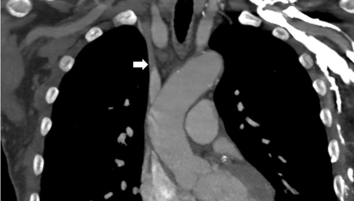
Figure 1
Computerized tomographic angiography demonstrating a type 3 thoracic central venous obstruction.
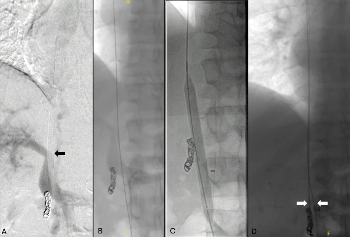
Figure 2
Intraoperative fluoroscopy images. (A) Angiogram demonstrated an uncrossable retrohepatic inferior vena cava occlusion. (B) Wire access was obtained into the right atrium via the left femoral venous catheter. (C) Balloon angioplasty of the inferior vena cava was then performed, (D) and a second wire advanced into the right atrium from the right groin.
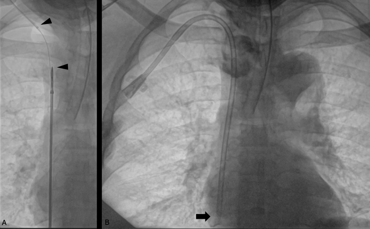
Figure 3
Fluoroscopy images showing (A) successful delivery of the surfacer device after recanalization of the retrohepatic inferior vena cava. (B) The tip of the tunneled dialysis catheter was then successfully placed in the right atrium.