
Introduction
Thoracic central venous obstructions (TCVOs) are a common complication of central venous catheter insertions in patients on dialysis.1 The Surfacer® Inside-Out® (SIO) Access Catheter System from Merit Medical enables right-sided central venous placement in patients presenting with TCVOs who have undergone failed attempts at catheterization or recanalization through conventional means.2 The device allows for an inside-out approach through right femoral access.1, 3 Reported success rates range from 88% to 100%,4 although this technique is generally contraindicated in patients with acute iliocaval thrombosis.5 In this case report, we demonstrate the successful placement of the SIO device through an occluded inferior vena cava.
Case Presentation
Clinical Presentation
A 65-year-old male with a prior medical history of hypertension and end-stage renal disease on hemodialysis with a right upper extremity Hemodynamically Reliable Outflow (HeRO®) graft by Merit Medical presented to our institution due to a HeRO graft malfunction. He had a known central venous occlusion with multiple prior arteriovenous access failures. His HeRO graft, which had been placed 5 years prior to admission, was complicated by graft infection with a recent hospitalization for bacteremia. The patient now had a left femoral tunneled catheter for dialysis. On the physical exam, the right upper extremity HeRO graft material was exposed and nonfunctional. Distal pulses in the radial and ulnar arteries were palpable and symmetric bilaterally. No externalized bleeding or edema was noted. The exposed graft was then explanted at bedside, and a computerized tomographic venogram was obtained for further access planning.
Preoperative Imaging and Planning
Preoperative computerized tomography (CT) venogram revealed occlusion of the bilateral brachiocephalic veins extending from the superior vena cava confluence to the left subclavian and internal jugular veins, thus demonstrating a type 3 TCVO (Figure 1). Given that this patient required urgent permanent arteriovenous access for dialysis, the plan was to establish a right-sided central access using the SIO technique and subsequently convert to another HeRO graft.
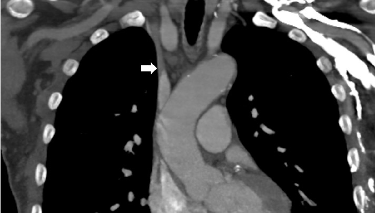
Figure 1
Computerized tomographic angiography demonstrating a type 3 thoracic central venous obstruction.
Operative Details
The patient was brought to the hybrid room for an SIO procedure as planned. We obtained ultrasound-guided access of the patent common femoral vein in the right groin using a micropuncture kit. The prior left groin venous catheter remained in place for planned contrast injection during on-table cone-beam CT angiography (CBCTA) as previously described in the literature.6 A Bentson wire was advanced, and the access was upsized to a 12F sheath. We then performed a cavogram, which demonstrated an occluded retrohepatic inferior vena cava (IVC) (Figure 2A).
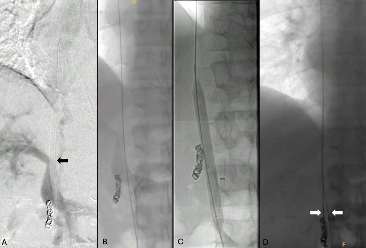
Figure 2
Intraoperative fluoroscopy images. (A) Angiogram demonstrated an uncrossable retrohepatic inferior vena cava occlusion. (B) Wire access was obtained into the right atrium via the left femoral venous catheter. (C) Balloon angioplasty of the inferior vena cava was then performed, (D) and a second wire advanced into the right atrium from the right groin.
Several attempts to cross the occlusion were unsuccessful. Since this patient had a left-sided femoral catheter with the tip lying beyond the IVC occlusion, we successfully delivered an Amplatz wire (Boston Scientific) into the right atrium through the catheter (Figure 2B). The catheter was then easily pulled out and exchanged for a 12F sheath. Once access was secured, an 8 × 8 mm Conquest™ balloon (Becton Dickinson) was advanced over the wire, and angioplasty of the lesion was successfully performed (Figure 2C) with minimal residual stenosis. Next, a combination of a Bern catheter (Boston Scientific) and a stiff-angled GLIDEWIRE® (Terumo Medical Corporation) was used to cross the retrohepatic IVC from the right groin to secure the right access.
We then proceeded with the SIO technique as intended. The Surfacer workstation sheath was inserted through the right groin, after which the device was introduced and advanced to the tip of the sheath. Next, according to our institutional practice, an on-table CBCTA was performed to ensure that no arterial structures were in the projected trajectory of the device. Upon confirmation, the device was carefully advanced through the TCVO and the needle wire deployed toward the skin and externalized. A 5F sheath was then placed over the externalized wire into the SVC (Figure 2D), and a pullback venogram was performed to ensure no contrast was seen in the arterial system or in the pleural cavity.
Next, a 23-cm tip-to-cuff tunneled dialysis catheter was brought onto the field. An 11 blade was used to create a skin incision at the access point and the intended exit site of the chest wall. The catheter was then tunneled appropriately. Next, a peel-away sheath was introduced over the externalized wire, and the TDC was delivered into the right atrium through the peel-away sheath. The tip of the catheter was confirmed on x-ray to be in the mid-right atrium (Figure 3) with excellent flushing and blood return. The ports were packed with heparin and all the access devices were removed and pressure held to obtain hemostasis. The catheter was affixed to the chest wall using a 2-0 nylon suture, and the neck access point was closed using a 4-0 MONOCRYL™ suture (Ethicon US, LLC). The patient tolerated the procedure well without complications and was taken to recovery in stable condition.
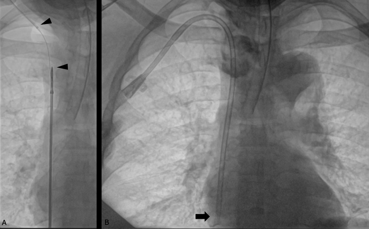
Figure 3
Fluoroscopy images showing (A) successful delivery of the surfacer device after recanalization of the retrohepatic inferior vena cava. (B) The tip of the tunneled dialysis catheter was then successfully placed in the right atrium.
Postoperative Course
The immediate postoperative course was notable for bleeding from the right groin access site with marked hypotension and cardiac arrest while the patient was heading home after discharge. Spontaneous circulation returned after one round of cardiopulmonary resuscitation, and the patient was admitted to the intensive care unit. His in-hospital stay was uneventful, and he was discharged home on postoperative day 4. At his 30-day follow-up, the patient was doing well without any active issues with his catheter, which was successfully converted to a HeRO graft 4 months later.
Discussion
This case demonstrates successful delivery of the SIO Access Catheter System through an occluded IVC. The contraindication of using SIO in IVC occlusions stems from issues with obtaining safe access to the thoracic central venous system. This is especially true in acute iliocaval thrombosis, where there is increased risk of dislodging the clot into the right atrium, with potential for a pulmonary embolism.6, 7 Also, there have been no previous trials to evaluate the safety of the SIO procedure in patients with iliocaval obstructions. In our patient, however, the IVC occlusion appeared chronic, within the context of an indwelling catheter. We therefore deemed it safe to attempt recanalization of the lesion to deliver the surfacer device. While this is admittedly an off-label use of the device, it must be noted that patients who require an SIO procedure generally have exhausted all alternative upper extremity access options and therefore usually require unconventional means, such as described in this article, to ensure they obtain adequate access for hemodialysis.
In this case, the presence of an indwelling catheter offered an ingenious recanalization pathway through the lesion, making it possible to dilate the track and deliver the device through the right groin.8 Despite a successful procedure, our patient presented an unexpected serious bleeding complication, which was deemed not to be device related. Indeed, groin bleeding is a common complication of endovascular procedures.9, 10, 11 warranting effective access closure and close monitoring of patients in the immediate postoperative period for early detection to prevent catastrophic consequences.
Conclusion
The SIO procedure has proven to be an invaluable option for central venous catheterization in patients who have exhausted all access options in the chest and upper extremities; however, it does come with its own anatomic restrictions. This case demonstrates that delivery of the device through a chronically occluded inferior vena cava is feasible once a recanalized path has been achieved. Further studies are warranted to investigate the safety and effectiveness of this technique.
Competing Interests
Dr. Peden was a consultant for Merit Medical and a principle investigator in the Investigational Device Trial for FDA approval of the Surfacer Device. He also is currently a consultant for Venostent, Voyager, and Humacyte Inc. All fees are retained by Houston Methodist Hospital. Rishik Puppala, Pallavi Gorantla, and Bright Benfor have no conflicts to disclose.