
Introduction
Heart failure is a progressive and eventually terminal disease that impacts communities and societies worldwide.1,2 Many causes can contribute to heart failure, including coronary artery disease,3 hypertension,4 obesity,5,6 diabetes mellitus, sarcoidosis, amyloidosis, structural heart disease,7 infectious etiologies (ie, viral, parasitic or others),8 toxins (alcohol, cocaine, methamphetamine, etc.),9 muscular dystrophies, and many others.10,11 Seminal studies have demonstrated benefits and improved survival using reverse remodeling agents/medications in patients with heart failure. Such guideline-directed medical therapies include beta adrenergic receptor blockers, mineralocorticoid receptor antagonists, renin-angiotensin system inhibitors (angiotensin receptor-neprilysin inhibitors, angiotensin converting enzyme inhibitors, angiotensin receptor blockers), and sodium-glucose cotransporter inhibitors.12 In combination with these medical therapies, device therapies such as cardiac resynchronization therapies (cardiac resynchronization therapy or biventricular pacing) or implantable cardioverter-defibrillators have been shown to improve survival in patients with severe, end-stage heart failure. Despite these major advances using medical and device therapies, 50% of patients with heart failure do not survive beyond 5 years from their initial date of diagnosis.12,13 The only curative therapy for end-stage heart failure is orthotopic allogeneic heart transplantation.14 In this brief review, we will examine this modality as well as alternative strategies and donor organ sources that may complement the use of allogeneic organ transplantation. We also highlight the current state and areas of investigation related to cardiac transplantation that are underway in order to broadly apply these therapies to patients with end-stage cardiovascular diseases.
Allogeneic Cardiac Transplantation
The first orthotopic allogeneic heart transplant was performed in Cape Town, South Africa (Groote Schuur Hospital), by Christiaan N. Barnard in 1967.15 Initially (1967-1980), patient survival following allogeneic heart transplantation was limited. However, scientific advances in immunobiology transformed the field and improved the survival of transplant recipients. Specifically, the discovery of cyclosporine in 1972 and its eventual approval by the US Food and Drug Administration for use as an immunosuppressant agent in 1983 improved graft and patient survival.16 Today, most immunosuppression protocols include a corticosteroid taper, a reversible inhibitor of inosine monophosphate dehydrogenase (mycophenolate mofetil), and a calcineurin inhibitor (tacrolimus or cyclosporine).17
Worldwide, more than 5,000 heart transplants are performed each year and have a 10-year survival of approximately 50%.18 While intense efforts remain focused on immunological barriers and the long-term impact of immunosuppression, allogeneic heart transplantation remains the only curative therapy for end-stage heart failure. However, the limited number of donor organs precludes the broad application of solid organ transplantation for end-stage (terminal) heart disease. Moreover, it is estimated that as many as 100,000 to 500,000 individuals worldwide could benefit from such therapy.18 This discrepancy between those individuals who need an organ transplant and those who receive such lifesaving therapies is what drives the pursuit of alternative organ sources.
Xenotransplantation and the Use of Gene Edited Organs
The limited supply of donor organs has ignited a longstanding interest and investigation focused on xenotransplantation. The first cardiac xenotransplant was performed in 1964 when an undersized chimpanzee heart was transplanted in a patient with cardiogenic shock and was immediately rejected.19,20,21 Approximately 20 years later, a baboon heart incompatible with the ABO blood type was transplanted into a newborn patient (Baby Fae) who had hypoplastic left heart syndrome (failure) and survived for approximately 20 days.19,20,21,22 These pioneering initiatives provided an important basis for further studies and the engineering of improved xenografts.
The development and use of gene editing technologies in combination with somatic cell nuclear transfer technology provided new opportunities to engineer pig organs for xenotransplantation. The pig is an attractive xenograft source for many reasons as it has a rapid growth phase to physical maturity and has a comparable size, anatomy, and functional (physiological) performance compared to the human heart. Therefore, efforts have further focused on genetic modifications of swine as xenogeneic organ donors (Figure 1).
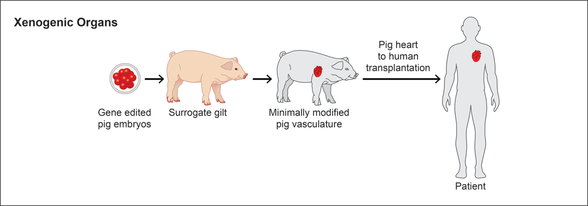
Figure 1
Genetically engineered pigs as organ sources for xenotransplantation. Using gene disruption and transgenic technologies, juvenile and adult pigs have been engineered and serve as organ sources for xenotransplantation.
There is broad consensus that the most prevalent carbohydrate antigen on pig cells, galactose-α(1,3)-galactose (αGal), should be eliminated since it is recognized by large amounts of preformed natural antibodies in all humans and Old World nonhuman primates (ie, several species of monkeys found in Africa and Asia such as baboons, macaques, etc.).23,24 Many but not all investigators consider other carbohydrate targets of natural antibodies, including N-glycolylneuraminic acid, and a human Sd(a) blood group-like glycan, also important molecules to be eliminated.25,26 Binding of antibodies to these targets leads to activation of complement and coagulation cascades and antibody-mediated rejection of the xenotransplant. This problem can be overcome by inactivating the genes responsible for these antigens: GGTA1 (coding α-1,3-galactosyltransferase), CMAH (coding cytidine monophosphate-N-acetylneuraminic acid hydroxylase), and B4GALNT2/B4GALNT2L (coding β-1,4-N-acetyl-galactosaminyl transferase 2).26 Cells from these triple-knockout (TKO) pigs show very little human natural antibody binding, whereas baboons have more antibodies against TKO pig cells than to GGTA1 knockout cells.27
A complementary strategy is the expression of one or more human complement pathway regulatory proteins (such as human CD46), which block different steps in the activation of the complement cascade. By combining these strategies, hyperacute rejection of pig-to-primate xenografts can be completely controlled.27
Another important issue is the dysregulation of coagulation in the xenotransplanted heart, eventually leading to thrombotic microangiopathy and graft failure. This problem can be mitigated by transgenic expression of human coagulation regulating proteins. Thus far, the most effective approach has been the expression of human thrombomodulin (hTHBD) that, in complex with human thrombin, activates protein C, resulting in a strong anticoagulant function.28 Indeed, hearts from triple-modified pigs (genetically knocking out of αGal and transgenic expression of proteins hCD46 and hTHBD) survived consistently for up to 6 months after orthotopic transplantation into baboons.29,30,31 In addition to genetic modification of the source pigs, cold nonischemic perfusion of the donor hearts, nontoxic immunosuppression of the recipients using CD40-CD154 costimulation blockade, and post-implantation growth control of the xeno-heart using the mechanistic target of rapamycin inhibitor temsirolimus were key to this success.32
Additional transgenes, including other complement pathway regulators, human endothelial protein C receptor, human heme oxygenase I, or human CD47 did not provide a significant advantage over this basic combination of a GGTA1 knockout plus expression of hCD46 and hTHBD.33 An additional modification associated with an even longer survival (9 months) in baboons was the knockout of the growth hormone receptor gene (GHR) to prevent excessive growth of the donor heart (although the GHR KO pig heart following xenotransplantation does continue to grow albeit at a slower rate).33,34 However, GHR KO pigs show several alterations, including transient juvenile hypoglycemia, marked obesity, and changes in liver metabolism.35,36
Selection of a genetic background of source pigs, such as miniature swine,37,38,39 which are already the same size as humans, seems preferable. In this case, TKO plus expression of hCD46 and hTHBD may be a sufficient set of genetic modifications for clinical xenotransplantation trials,27,32 although source pigs with much more complex modifications have been generated (such as genetically modified pigs with four genes that have been deleted and transgenic expression of six human proteins).40,41 However, multiple genetic modifications may complicate the breeding of the source pigs and suffer from unexpected new xenoantigens as well as interactions between transgenes or transgene products.42
Successful Nonhuman Primate Studies Provided an Important Foundation for the Transition to Human Patients
The world’s first and second cardiac porcine-human xenotransplantation was undertaken in 2022 and 2023 at the University of Maryland. Separately, two patients with terminal end-stage heart failure and significant comorbidities who were not candidates for other medical or surgical therapies received a porcine cardiac xenograft from a genetically modified pig. These patients received intense immunotherapies and survived up to 2 months following the procedure.40,43,44 While these efforts were successful at demonstrating the feasibility and prevention of hyperacute rejection, these initiatives emphasized the importance of performing a clinical trial in well-evaluated and selected recipients to evaluate the efficacy of xenotransplantation. Moreover, longer-term survival will be necessary before broad application of xenotransplantation can be introduced.
Furthermore, other issues require ongoing investigation, such as the imaging studies that are required to accurately size match the donor xenograft (heart) with the recipient to preclude xenograft failure. In addition, ongoing efforts are focused on the prevention of xenozoonoses by establishing closed swine herds within barrier facilities and routine surveillance screening for more than 100 pathogens (such as porcine cytomegalovirus, porcine herpesvirus, porcine coronaviruses, Hepatitis E virus, and others).2,31,32 In addition, gene editing has been used to disrupt the pol gene of porcine endogenous retroviruses (PERVs) although no PERV transmission has been detected in nonhuman primate recipients of porcine xenografts.2,32 These surveillance measures of potential pathogens involving the donor xenografts and the recipients will be important as the field of xenotransplantation continues to mature.
Exogenic Pig Organs for Transplantation
An additive concept to make xeno-hearts even more compatible would be the humanization of heart structural components, such as the endothelium.45 This approach, called exogenesis, is based on the formation of interspecies chimeras from host embryos that are genetically engineered to be deficient in the generation (or survival) of one or multiple lineages, thereby creating a niche(s) for donor cells of another species to replace the missing cells (or even organs) (Figure 2).46,47,48 For example, seminal studies were undertaken in two evolutionary-related species to engineer rat-mouse chimeras.49 In this study, Pdx1 null mouse embryos (which completely lacked a pancreas) were complemented at the blastocyst stage by injecting green fluorescent protein (GFP)-labelled rat embryonic stem cells (ESCs). The resulting viable interspecies chimeras developed a normal functioning GFP-labelled rat pancreas in the Pdx1 null recipient. Conversely, in the reverse experiments, Pdx1 null rat embryos injected with GFP-labelled mouse ESCs developed functional rat-sized mouse pancreases. To assess functionality, islets from these mouse-derived pancreases were transplanted into streptozotocin-induced diabetic mice of the ESC-donor strain. Remarkably, the transplanted islets maintained normal host blood glucose levels for over 370 days without immunosuppression.50 These findings provide proof-of-concept evidence for the therapeutic potential of pluripotent stem cell (PSC)-derived organs generated through interspecies organogenesis for rejection-free transplantation.
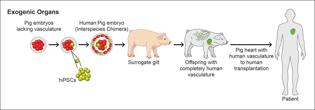
Figure 2
Exogenic organs using blastocyst complementation. Schematic highlighting the strategy for engineering exogenic, human organs in gene edited pigs. In this strategy, an entire lineage is deleted and the null porcine embryo is complemented by human induced pluripotent stem cells (iPSCs), which populate the vacant niche producing a human organ (ie, heart). Such a strategy could be used to engineer individual exogenic organs (from the patient’s hiPSCs) or an off-the shelf (human leukocyte antigens compatible hiPSCs) version.
Further studies have explored ways to enhance the efficiency of interspecies chimerism. For instance, introducing the anti-apoptotic gene BCL2 into PSCs or utilizing cell competition by employing IGF1 receptor-deficient hosts has been shown to improve chimerism efficiency.48,51,52 These investigations focus on key biological mechanisms, including apoptosis regulation, growth competition, and the development of a cell-competitive niche. These and other important landmark studies provide a foundation for engineering interspecies organs in the pig.
In contrast to the rat and mouse, pigs and humans are evolutionary more distant (ie, more than 90 million years separation). One of the challenges of evolutionary separation is that certain (divergent) species may have barriers for chimera formation of whole organs. The barrier is due to differences in development tempo, gene regulatory networks, and signalling pathways. Nevertheless, pig embryos (E18-19) with human endothelium have been generated by complementing pig blastocysts lacking ETV2, a master regulator of hematoendothelial lineages, with human induced pluripotent stem cells overexpressing the antiapoptotic factor BCL2.45
Another possibility to generate vasculogenesis-impaired porcine host embryos is to knockout the kinase insert domain receptor gene. Recently, hiPSC complementation of pig blastocysts with disrupted MYF5/MYOD/MYF6 genes has proved successful for the generation of growing porcine embryos with human muscle.53 Current attempts focus on the improvement of interspecies chimerism, eg, by inactivating the IGF1R gene in the host embryo.48 Systematic analyses of early porcine heart development should uncover new strategies for improving the efficiency of porcine blastocyst complementation with human stem cells. Another technically and even more demanding approach would be complementation of the respective organ niche at a later developmental stage in utero, or conceptus complementation.52,54 Overall, this approach has tremendous opportunities but will require enhanced understanding of developmental mechanisms and crosstalk between divergent species during organogenesis.
Conclusions
Heart failure is a devastating, terminal disease. The only curative therapy for end-stage heart failure is solid organ transplantation. While allogeneic heart transplantation has acceptable outcomes, this therapy is limited due to the shortage of donor organs. Alternative strategies include xenogeneic and exogenic organ production. Recent studies have demonstrated the feasibility of using genetically engineered porcine xenografts for transplantation into human patients. However, improvements will be required to obtain acceptable survival (≥ 1 year). Basic science studies support the notion that an exogenic approach could be an alternative strategy. Although the exogenesis approach has the potential to generate organs with better compatibility, there is one disadvantage: the whole procedure must be done for each individual organ (unless one is completely humanizing the vasculature), while complete xeno-hearts may be produced by breeding once the right genotype of source pigs has been established, especially if the animals are highly inbred, so that they are relatively uniform.37 Alternatively, the complete humanization of the endothelial lineage and/or vasculature may produce a pig that could be a source for all organs. Future studies will examine these hurdles and others with the aim of establishing a large donor source for patients with end-stage diseases such as heart disease.
Key Points
Allogeneic heart transplantation is a curative therapy for end-stage heart failure.
The advent of gene editing and somatic cell nuclear transfer technology provides an important platform to genetically modify source pigs for xenotransplantation.
Gene edited porcine xenograft organs have been successfully transplanted into patients with end-stage heart failure.
Gene editing and blastocyst complementation provide a platform for engineering exogenic organs.
Acknowledgements
The authors acknowledge the figure illustrations that were produced by Cynthia Faraday.
Competing Interests
Drs. Daniel J. Garry and Mary G. Garry are cofounders of NorthStar Genomics, LLC. Drs. Eckhard Wolf and Daniel Reichart are cofounders of XTransplant GmbH. David Sachs is a cofounder of Choironex and a Scientific Advisory Board member for ITB-MED. Hiro Nakauchi is a cofounder of Celaid Therapeutics, Ando Therapeutics, and Megakaryon Corp. Dr Joshua Weiner has no competing interest to declare.