
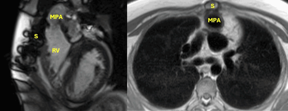
Figure 1
Cardiac magnetic resonance demonstrating right ventricle and main pulmonary artery adhesions to the sternum in a patient with double outlet right ventricle status post biventricular repair with arterial switch operation. S: sternum, RV: right ventricle, MPA: main pulmonary artery
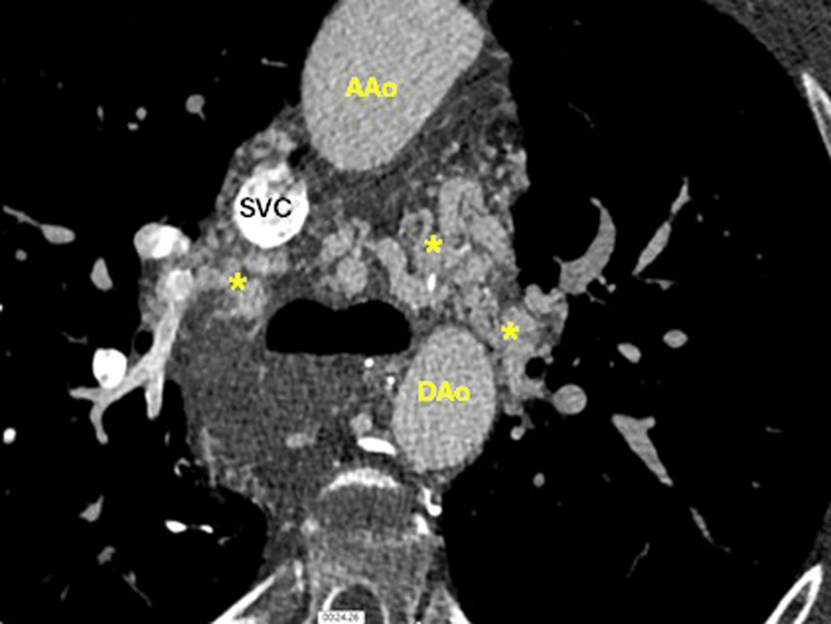
Figure 2
Computed tomography angiography of the chest with extensive collateral networks around the airway, the SVC and the aorta in a patient with unbalanced complete atrioventricular canal status post Fontan palliation. (*) Collateral networks, AAo: ascending aorta; Dao: descending aorta; SVC: superior vena cava
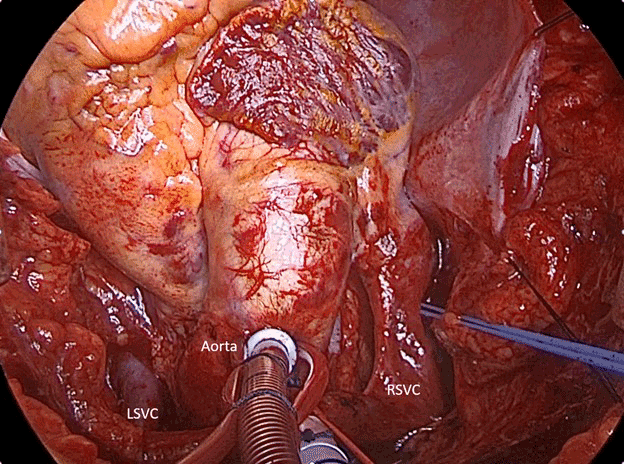
Figure 3
Patient with left superior vena cava (LSVC) and right superior vena cava (RSVC) who needs reconstruction for preparation for donor superior vena cava.
Video 1
Left superior vena cava (LSVC) reconstruction for Fontan patients who need superior vena cava; see also at https://youtu.be/KNPclQwR-UM.
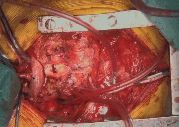
Figure 4
Patient who needs reconstruction of pulmonary arteries with a patch (*) to create a landing zone (*) for the donor pulmonary arteries.
Video 2
Pulmonary artery reconstruction; see also at https://youtu.be/zqP7aHtQ99Y.