
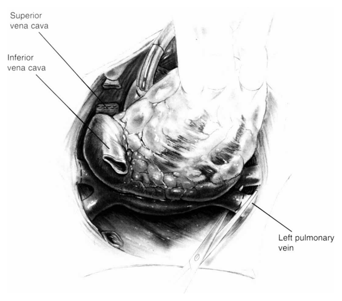
Figure 1
Shumway’s donor cardiectomy dividing the left atrial cuff. Reprinted from Morton Bolman R. Oper Tech Thorac Cardiovasc Surg. 1999 May;4(2):98-113. ©1999, with permission from Elsevier.
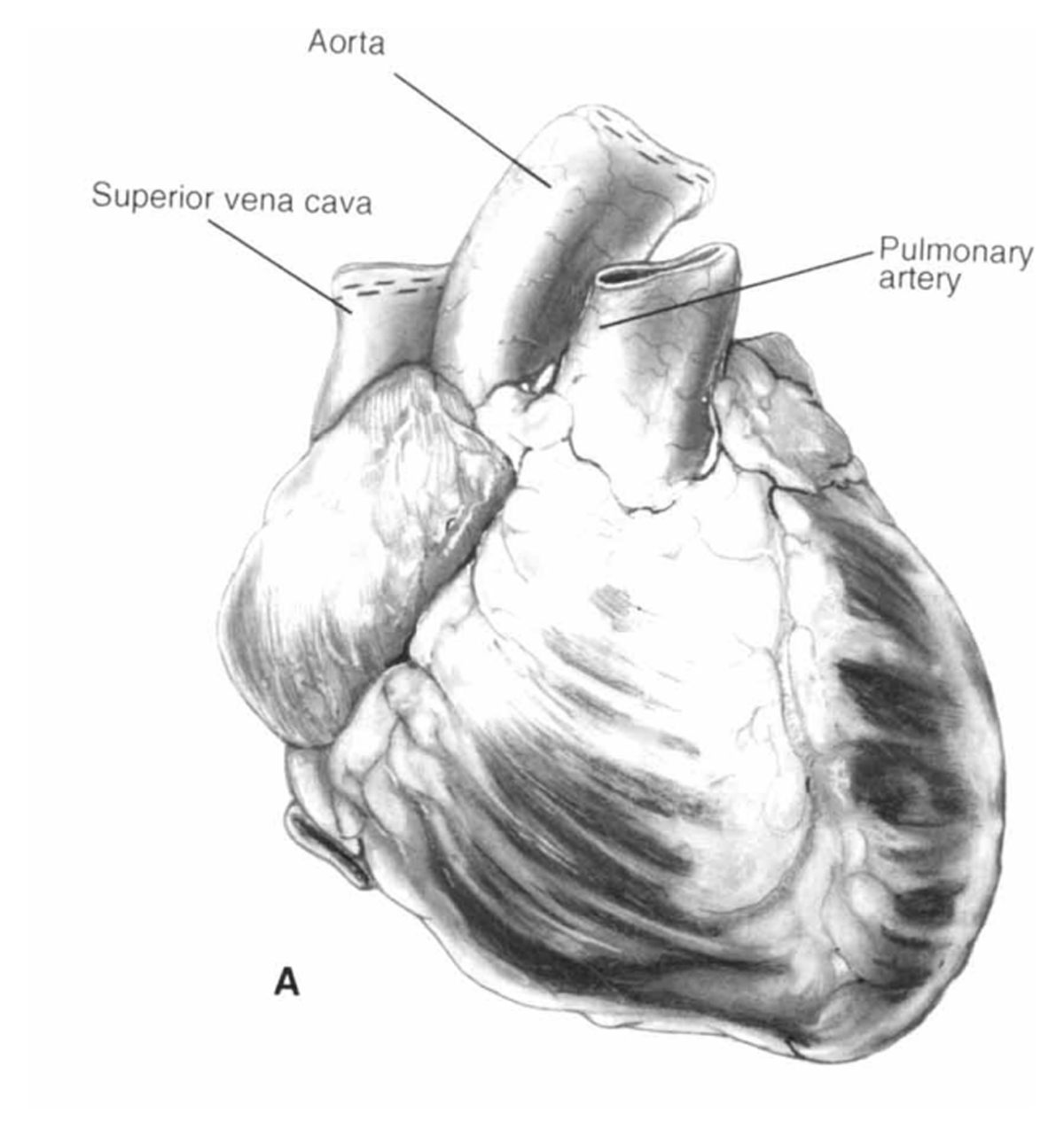
Figure 2
Shumway’s donor cardiectomy dividing the anterior vessels. Reprinted from Morton Bolman R. Oper Tech Thorac Cardiovasc Surg. 1999 May;4(2):98-113. ©1999, with permission from Elsevier.
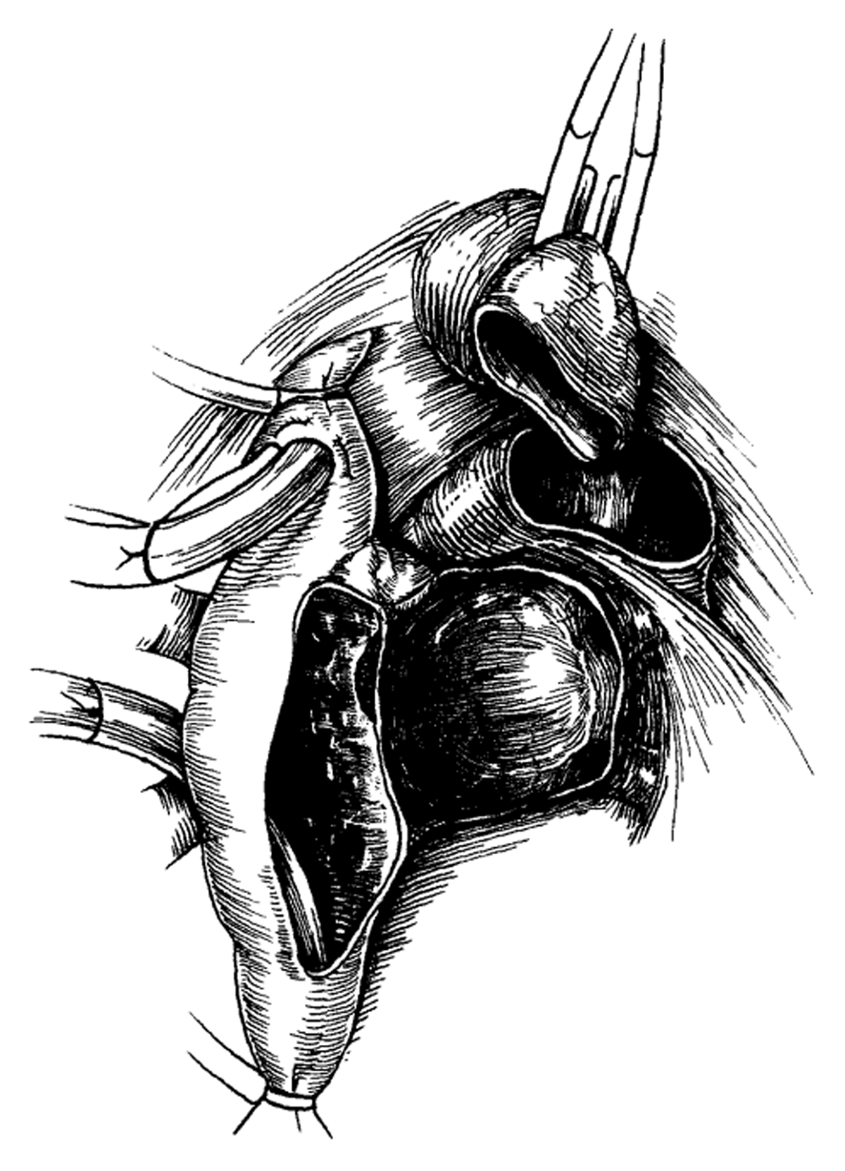
Figure 3
Biatrial technique completed for the recipient’s cardiectomy. Reprinted from Morton Bolman R. Oper Tech Thorac Cardiovasc Surg. 1999 May;4(2):98-113. ©1999, with permission from Elsevier.
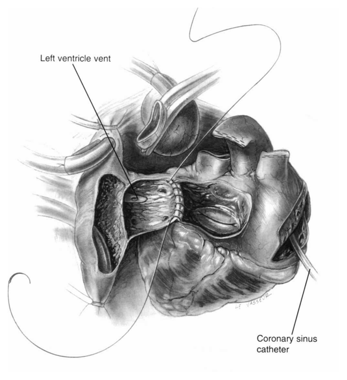
Figure 4
Biatrial technique using left atrial anastomosis. Reprinted from Morton Bolman R. Oper Tech Thorac Cardiovasc Surg. 1999 May;4(2):98-113. ©1999, with permission from Elsevier.
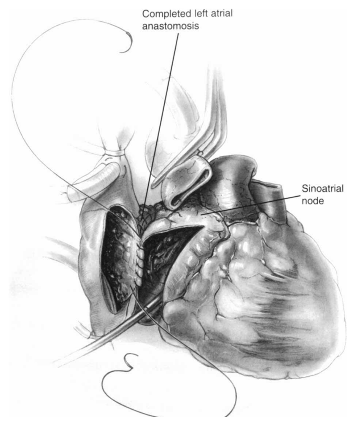
Figure 5
Biatrial technique using right atrial anastomosis. Reprinted from Morton Bolman R. Oper Tech Thorac Cardiovasc Surg. 1999 May;4(2):98-113. ©1999, with permission from Elsevier.
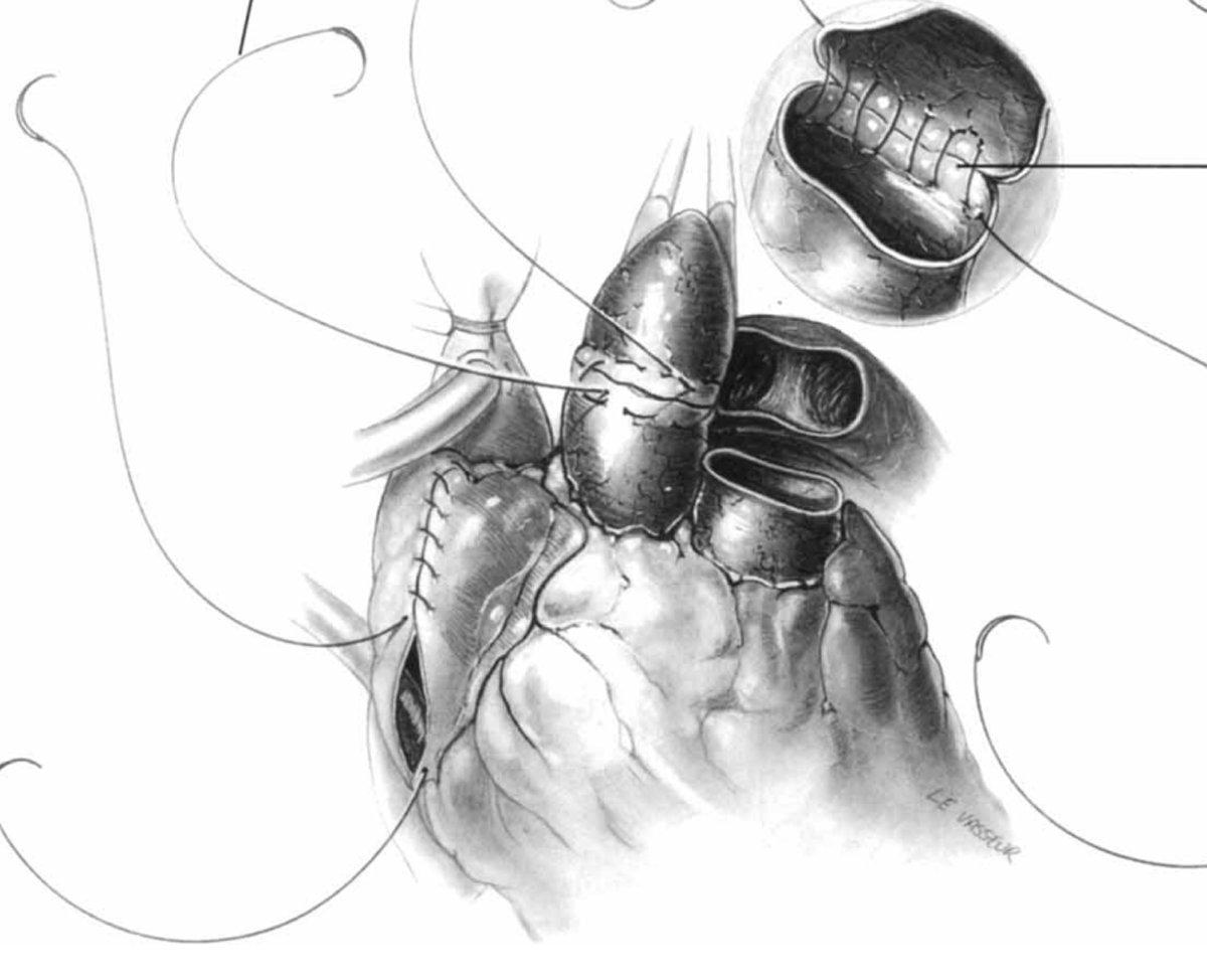
Figure 6
Pulmonary artery and aortic anastomoses used in biatrial technique. Reprinted from Morton Bolman R. Oper Tech Thorac Cardiovasc Surg. 1999 May;4(2):98-113. ©1999, with permission from Elsevier.
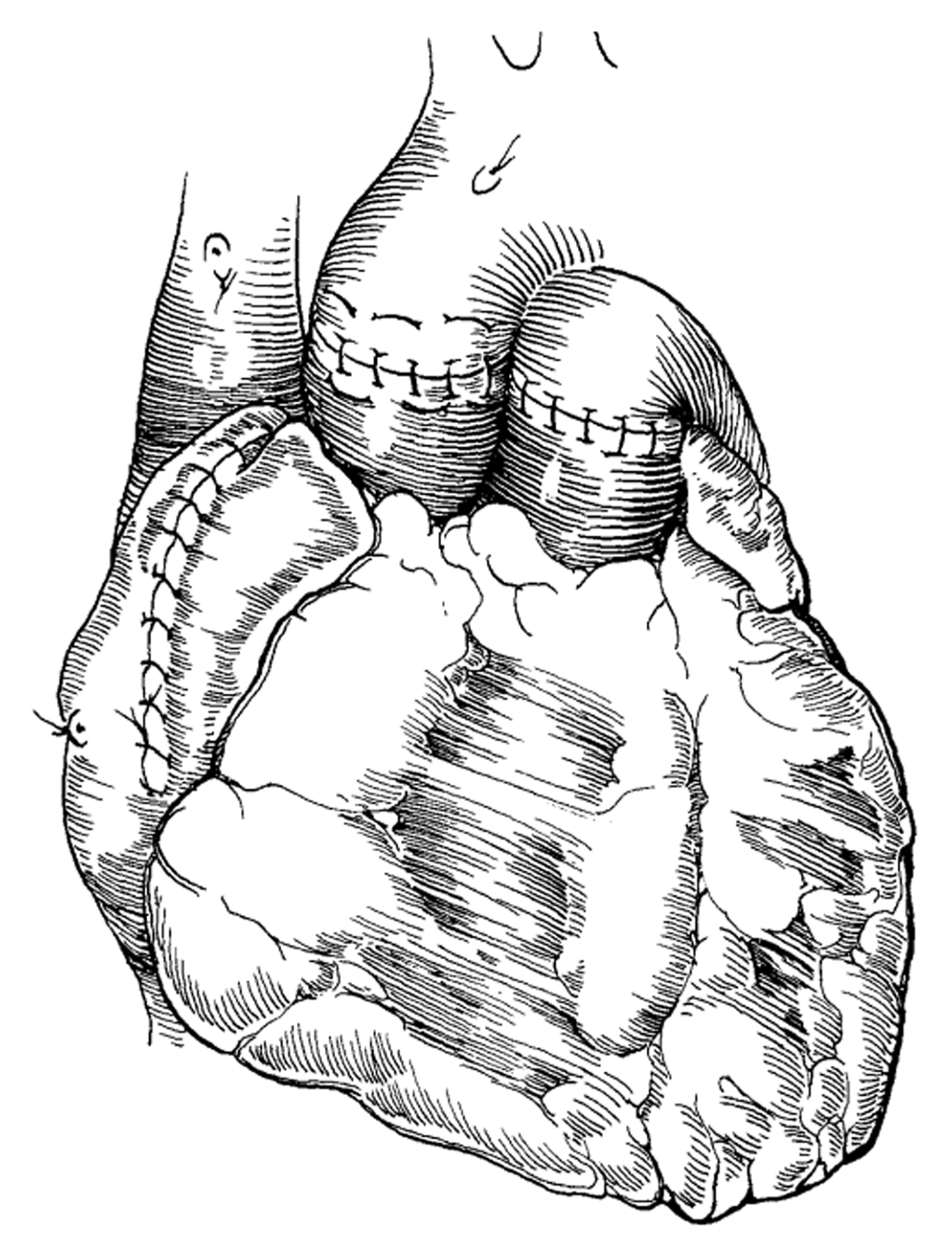
Figure 7
All the anastomoses are completed for the biatrial technique. Reprinted from Morton Bolman R. Oper Tech Thorac Cardiovasc Surg. 1999 May;4(2):98-113. ©1999, with permission from Elsevier.
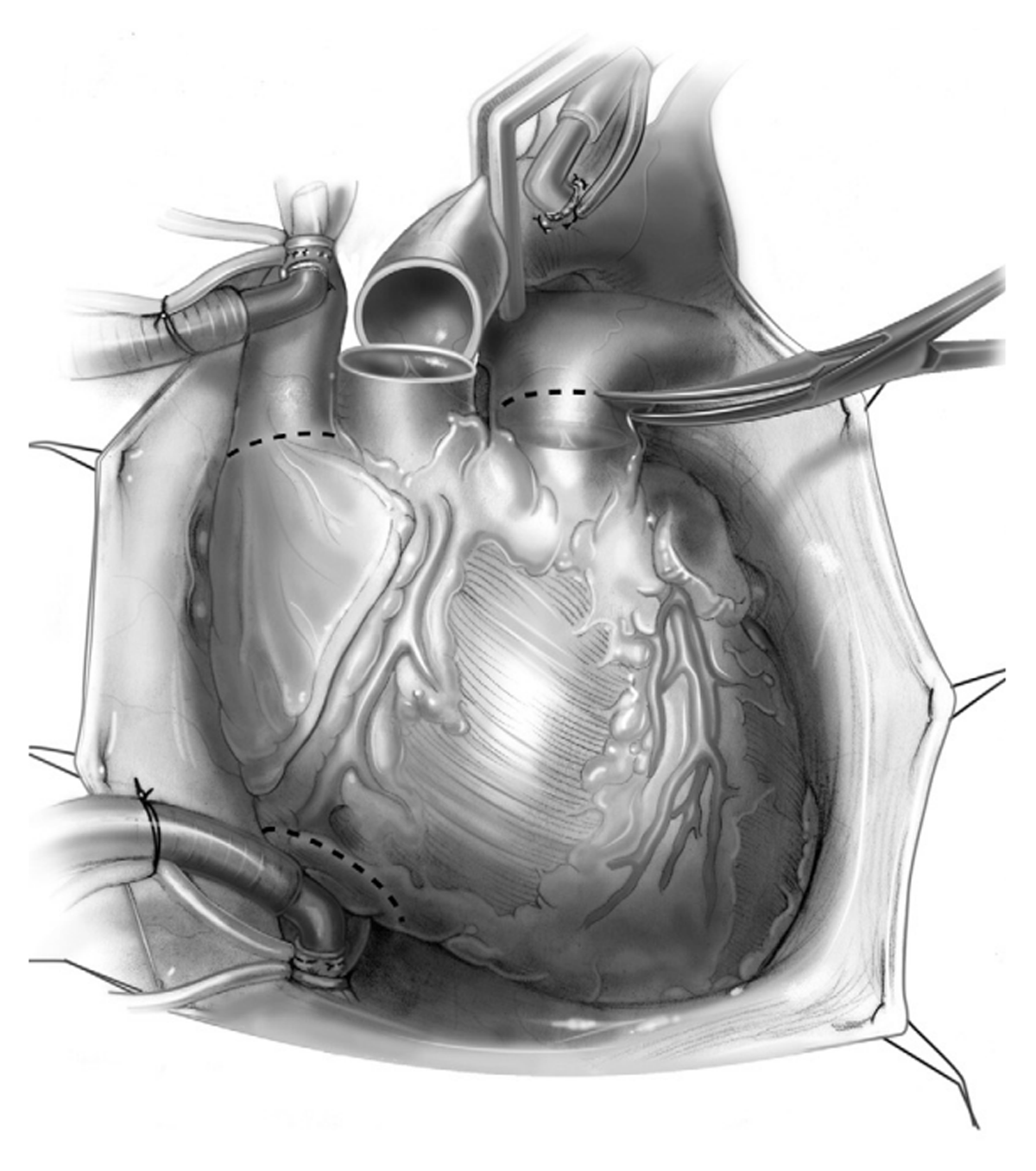
Figure 8
Donor cardiectomy used for bicaval implantation. Reprinted from John R, Liao K. Orthotopic Heart Transplantation. Oper Tech Thorac Cardiovasc Surg. 2010;15(2):138-146. ©2010, with permission from Elsevier.
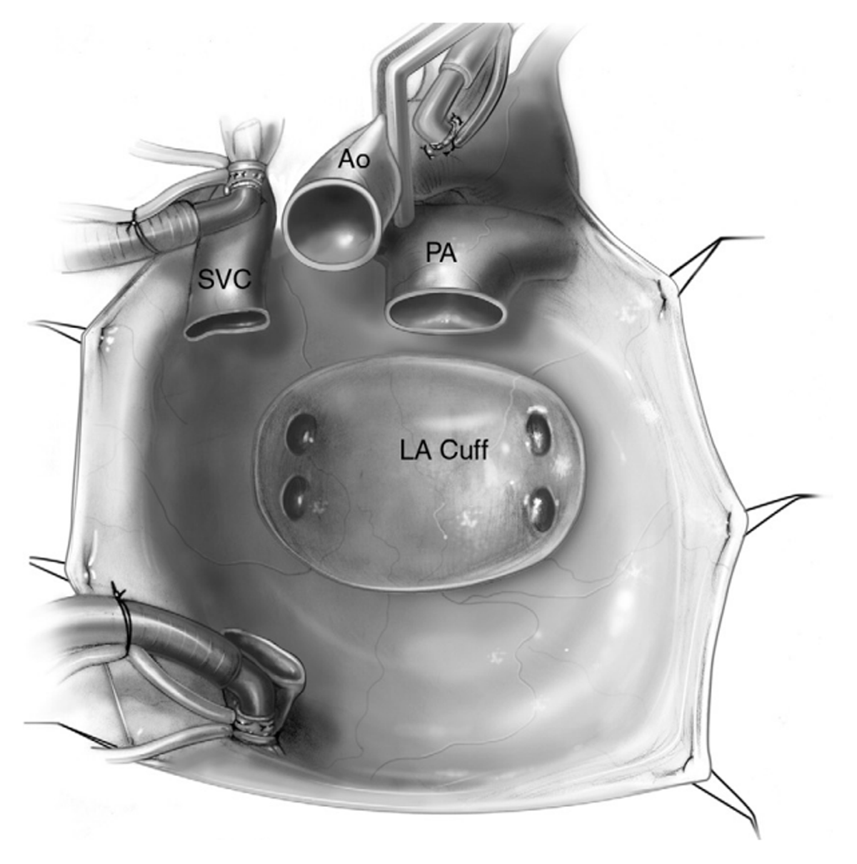
Figure 9
Recipient mediastinum after cardiectomy. Reprinted from John R, Liao K. Orthotopic Heart Transplantation. Oper Tech Thorac Cardiovasc Surg. 2010;15(2):138-146. ©2010, with permission from Elsevier. LA: left atrium; PA: pulmonary artery; Ao: aorta; SVC: superior vena cava
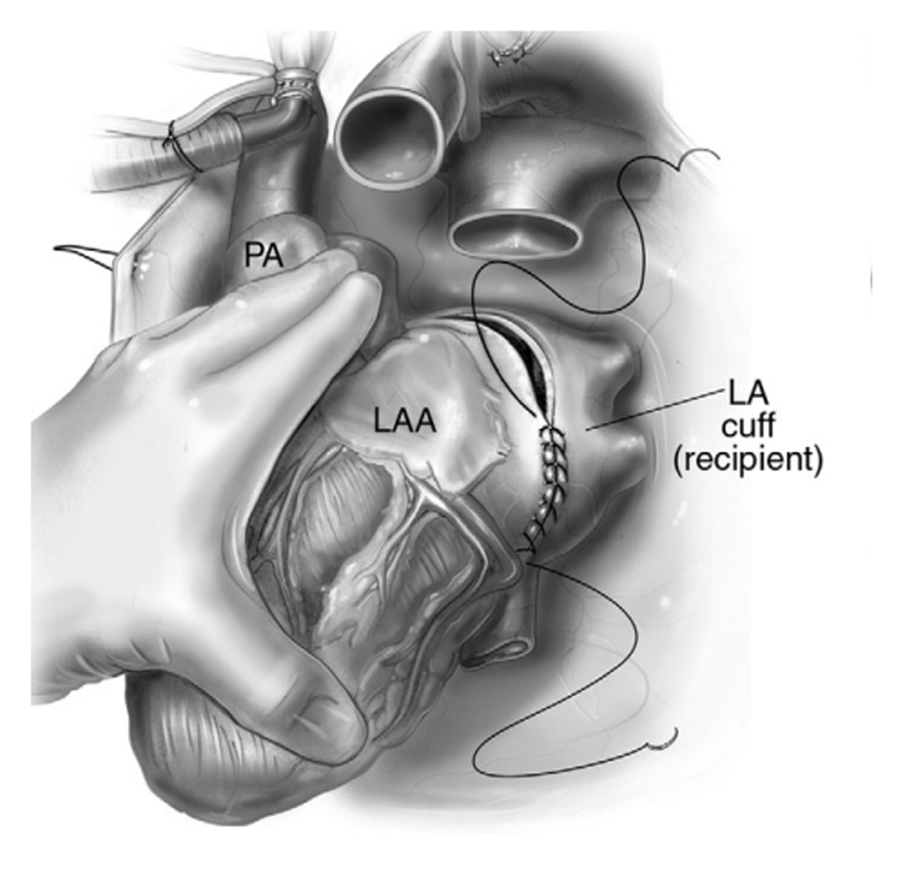
Figure 10
Bicaval technique using left atrial anastomosis. Reprinted from John R, Liao K. Orthotopic Heart Transplantation. Oper Tech Thorac Cardiovasc Surg. 2010;15(2):138-146. ©2010, with permission from Elsevier. LAA: left atrial appendage; LA: left atrium; PA: pulmonary artery
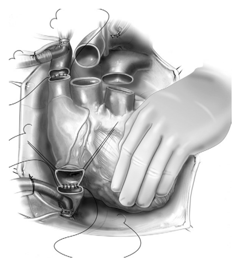
Figure 11
Inferior and superior vena cavae anastomosis used for bicaval technique. Reprinted from John R, Liao K. Orthotopic Heart Transplantation. Oper Tech Thorac Cardiovasc Surg. 2010;15(2):138-146. ©2010, with permission from Elsevier.
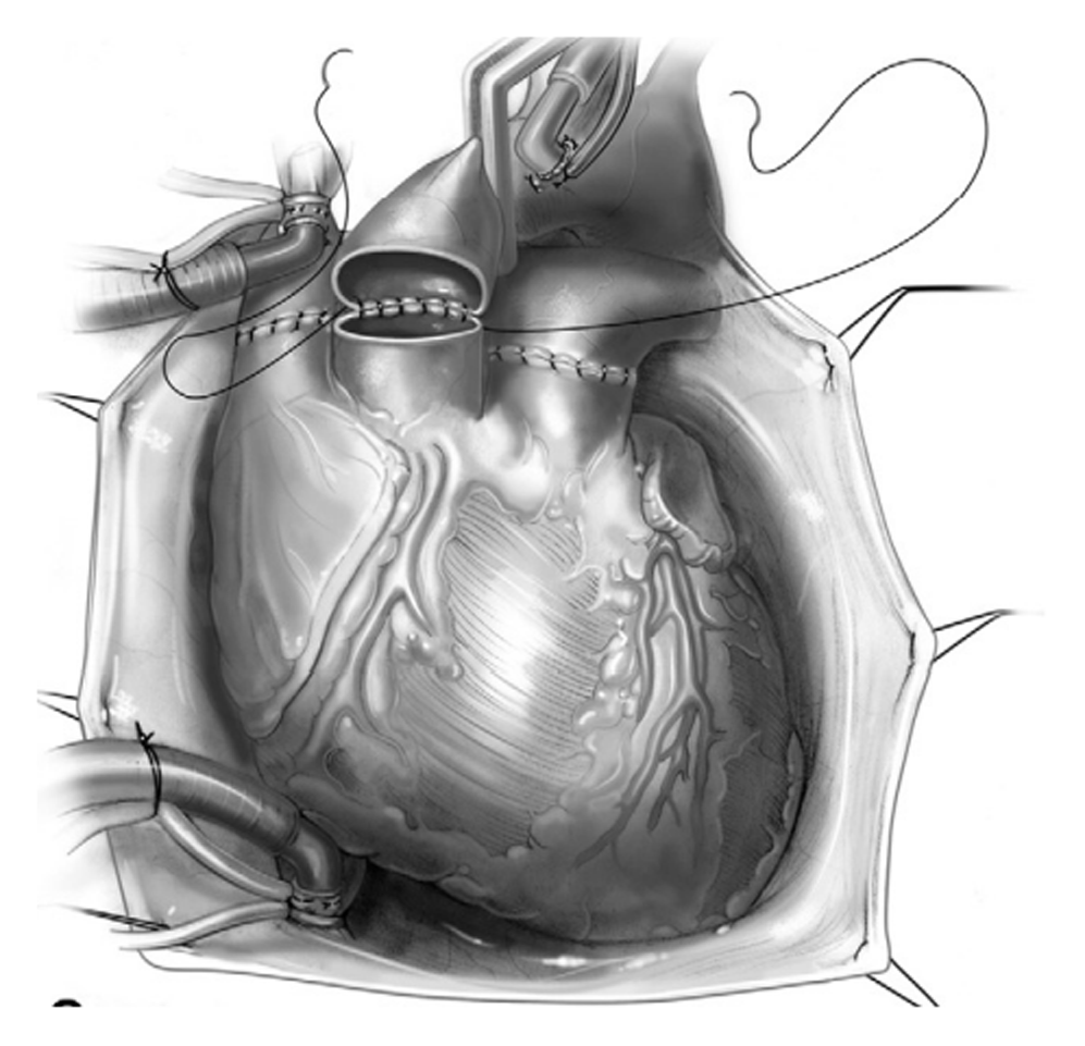
Figure 12
Aorta and pulmonary artery anastomosis used for bicaval technique. Reprinted from John R, Liao K. Orthotopic Heart Transplantation. Oper Tech Thorac Cardiovasc Surg. 2010;15(2):138-146. ©2010, with permission from Elsevier.
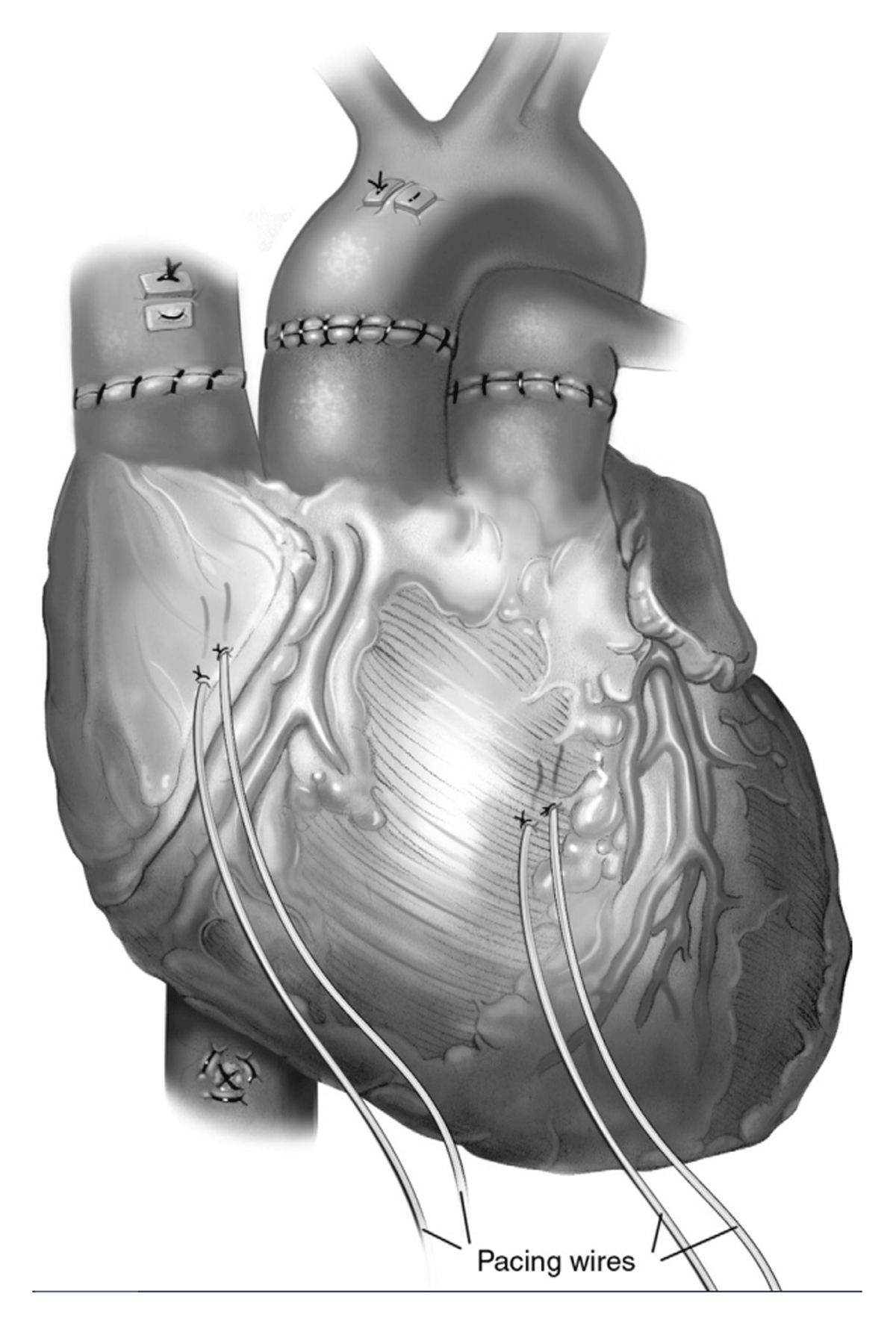
Figure 13
All the anastomosis are completed for the bicaval technique. Reprinted from John R, Liao K. Orthotopic Heart Transplantation. Oper Tech Thorac Cardiovasc Surg. 2010;15(2):138-146. ©2010, with permission from Elsevier.