
Introduction
Before the first human heart transplant occurred, studies on orthotopic homotransplantation of canine hearts were introduced by Richard Lower and Norman Shumway in the 1960s.
The first human heart transplantation was performed on December 3, 1967, by Christiaan Barnard in Cape Town, South Africa.1 Prior to this historic event, Barnard had accepted a scholarship at the University of Minnesota under Professor Wangensteen, where he met early pioneers of open heart surgery and the heart-lung machine. Barnard than returned to Groote Schuur Hospital and started an open heart surgery program to treat congenital defects and heart valve problems. A few years later, he visited the state of Virginia to learn immunosuppressive therapy. He also met Lower, who had been trained by Norman Shumway. Thus, Barnard gained experience with experimental heart transplantation.
His first transplant was performed on 54-year-old Louis Washkansky, who had coronary artery disease and was hospitalized due to acute heart failure related to ischemic cardiomyopathy. The donor heart came from a trauma victim with massive cerebral injuries, referred to as donation after circulatory death (DCD).
While the recipient was being prepared for transplantation, the donor’s heart was harvested using a technique originally developed by Shumway2 and was perfused mechanically at 10°C. Known as the biatrial technique for cardiac transplantation, this technique has been used in more than 20,000 patients. To prevent postoperative rejection, the recipient was given a cocktail of local irradiation, azathioprine, prednisone, and actinomycin C.
According to Shumway’s described technique, the donor’s left atrium is excised with the donor heart (Figure 1).3 Then the pulmonary veins are individually transected at the pericardial reflection. A cuff of left atrium is created by connecting incisions in these transected pulmonary veins, which depends on concomitant donor lung procurement. The superior vena cava (SVC) and inferior vena cavas (IVC) are divided, as are the aorta and pulmonary artery (Figure 2).3 This procurement technique has not changed since that time.
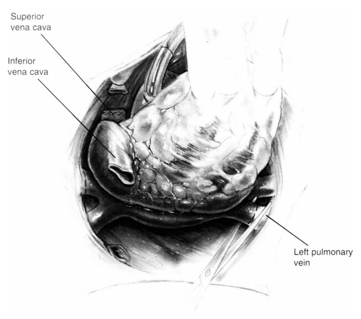
Figure 1
Shumway’s donor cardiectomy dividing the left atrial cuff. Reprinted from Morton Bolman R. Oper Tech Thorac Cardiovasc Surg. 1999 May;4(2):98-113. ©1999, with permission from Elsevier.
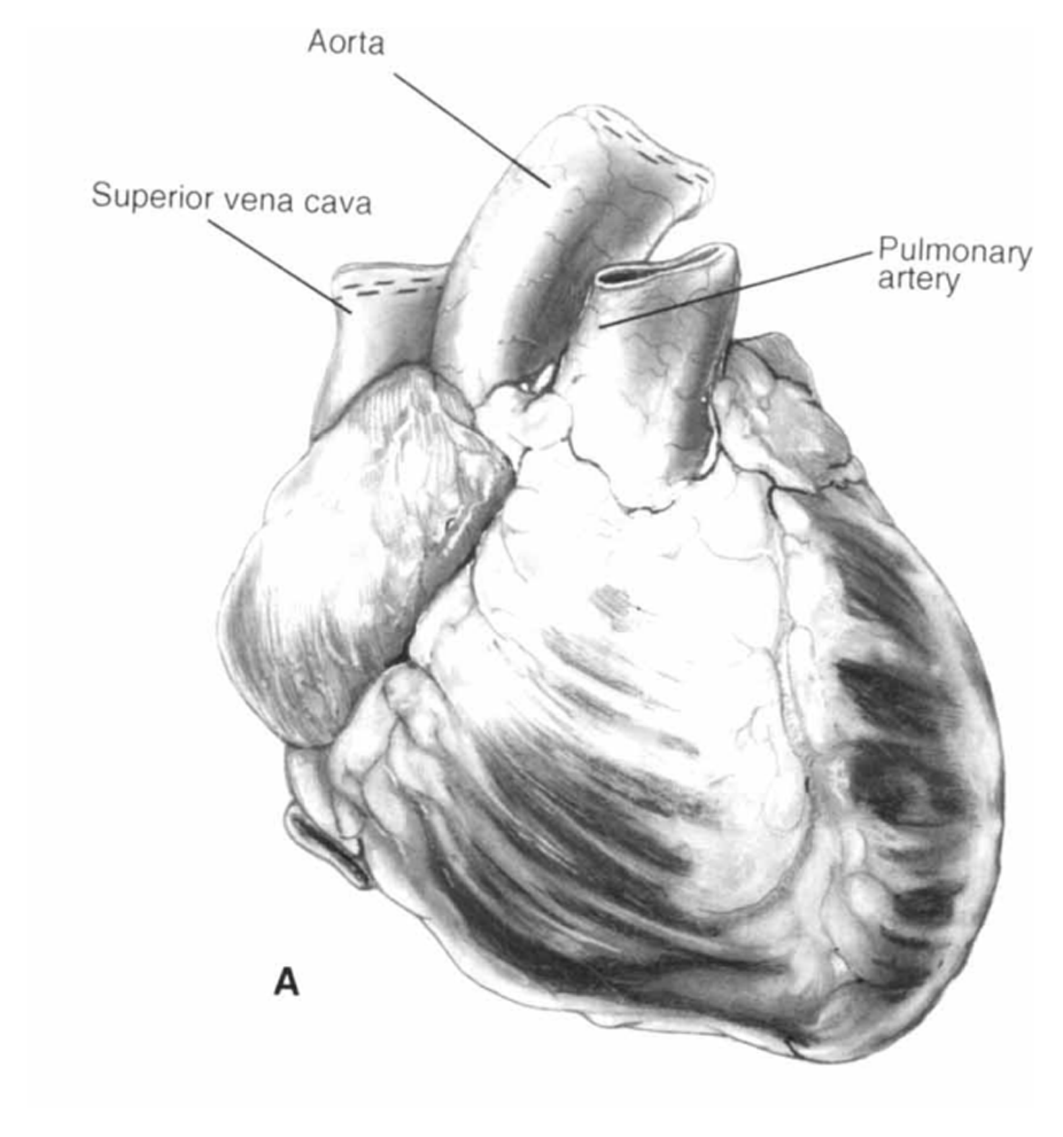
Figure 2
Shumway’s donor cardiectomy dividing the anterior vessels. Reprinted from Morton Bolman R. Oper Tech Thorac Cardiovasc Surg. 1999 May;4(2):98-113. ©1999, with permission from Elsevier.
Orthotopic Heart Transplant Implant Techniques
Historically, most of the early work on heart transplantation is attributed to Soviet Russian scientist Vladimir Demikhov, who performed a series of experimental operations on canines including orthotopic heart transplantation, heterotopic heart transplantation, and heart-lung transplantation en bloc.4 Since Demikhov’s studies were interrupted during World War II, he immediately returned to them in 1946 and performed canine intrathoracic transplantation of a heart, a lung, and the heart and lungs together without using a heart-lung machine. This first canine patient survived for 9.5 hours.
Demikhov created a closed-circuit circulation by clamping and dividing the thoracic aorta, IVC, brachiocephalic and subclavian arteries, and SVC. During transfer, the donor heart-lung was kept viable by its own closed-circuit circulation, with blood from the left ventricle being pumped into the arch of the aorta. From there it passed through the coronary vessels, supplying the myocardium, and then into the right atrium, right ventricle, and lungs. Oxygenated blood was returned to the left atrium. All this work and implantation technique was not known to the Western world until almost 20 years later, when D.K. Cooper’s historical paper in 1969 described Demikhov’s implantation technique.5
Biatrial Implantation Technique
The biatrial technique, which was refined and popularized by Lower and Shumway in 1960, was used to perform the first human heart transplantation.2
In this technique (Figure 3), the recipient’s cardiectomy is initiated by opening the right atrium along the atrioventricular groove where the coronary sinus demarcates the atrioventricular sulcus.3 The great vessels are transected above the semilunar commissures. The atria are incised along the atrioventricular grooves, leaving cuffs for allograft implantation. The stumps of the recipient aorta and pulmonary artery are suspended with stay sutures to retract these structures away from the field, thus facilitating exposure of the left atrial cuff for anastomosis.
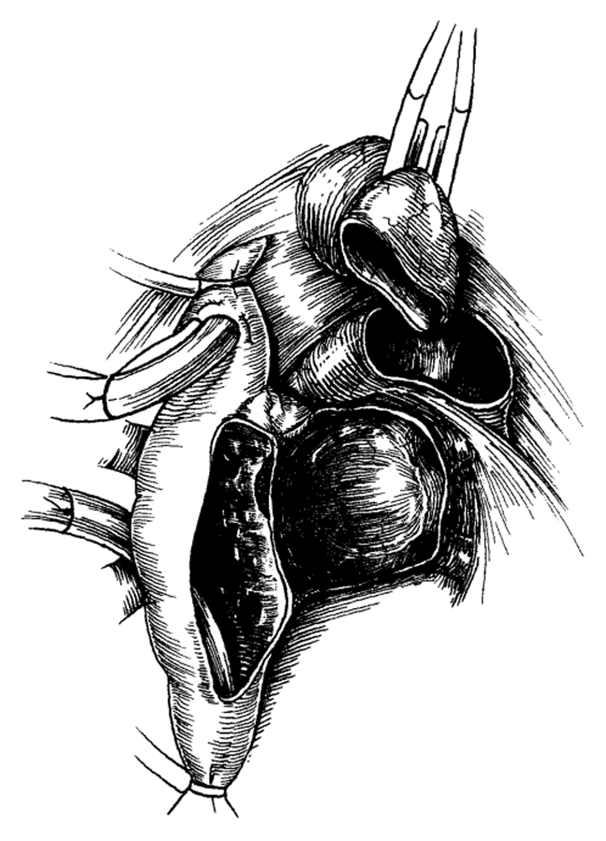
Figure 3
Biatrial technique completed for the recipient’s cardiectomy. Reprinted from Morton Bolman R. Oper Tech Thorac Cardiovasc Surg. 1999 May;4(2):98-113. ©1999, with permission from Elsevier.
Orientation of the donor heart to the recipient’s left atrial cuff is important. The sutures are passed multiple times, then the allograft is parachuted into the chest cavity. Left atrial anastomosis (Figure 4) is continued in a running fashion posteriorly and medially to the inferior aspect of the interatrial septum.3 Assessment of any size discrepancy is important in each suture between the donor and recipient; plication of access tissues might be necessary to achieve good hemostasis and anatomical geometry.
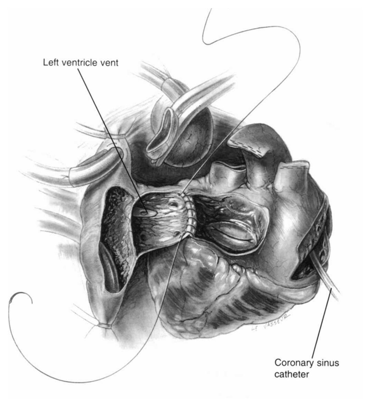
Figure 4
Biatrial technique using left atrial anastomosis. Reprinted from Morton Bolman R. Oper Tech Thorac Cardiovasc Surg. 1999 May;4(2):98-113. ©1999, with permission from Elsevier.
The right atrial anastomosis (Figure 5) is performed starting with the completed septal anastomotic site of the left atrial anastomosis and continuing to the already opened right atrial free wall in a running fashion.3
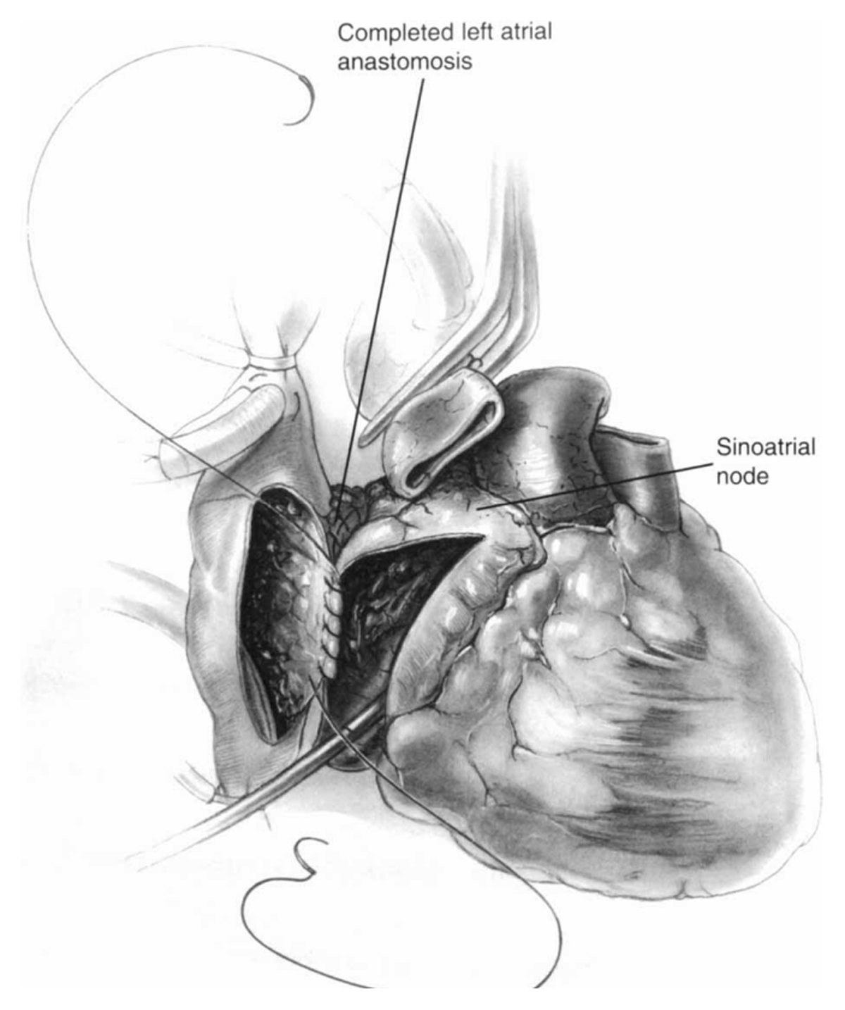
Figure 5
Biatrial technique using right atrial anastomosis. Reprinted from Morton Bolman R. Oper Tech Thorac Cardiovasc Surg. 1999 May;4(2):98-113. ©1999, with permission from Elsevier.
The end-to-end pulmonary artery anastomosis is performed (Figure 6).3 It is important to cut the pulmonary artery ends to an appropriate length to eliminate any redundancy in the vessel that may cause kinking, which is detrimental to right ventricular function. Finally, the aortic anastomosis is performed in a hemostatic fashion. Pulmonary artery and aorta anastomoses are not different for the biatrial orthotopic heart transplantation technique (Figure 7).3
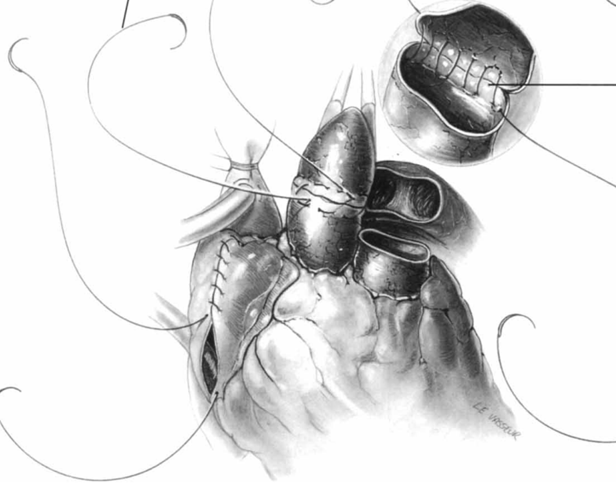
Figure 6
Pulmonary artery and aortic anastomoses used in biatrial technique. Reprinted from Morton Bolman R. Oper Tech Thorac Cardiovasc Surg. 1999 May;4(2):98-113. ©1999, with permission from Elsevier.
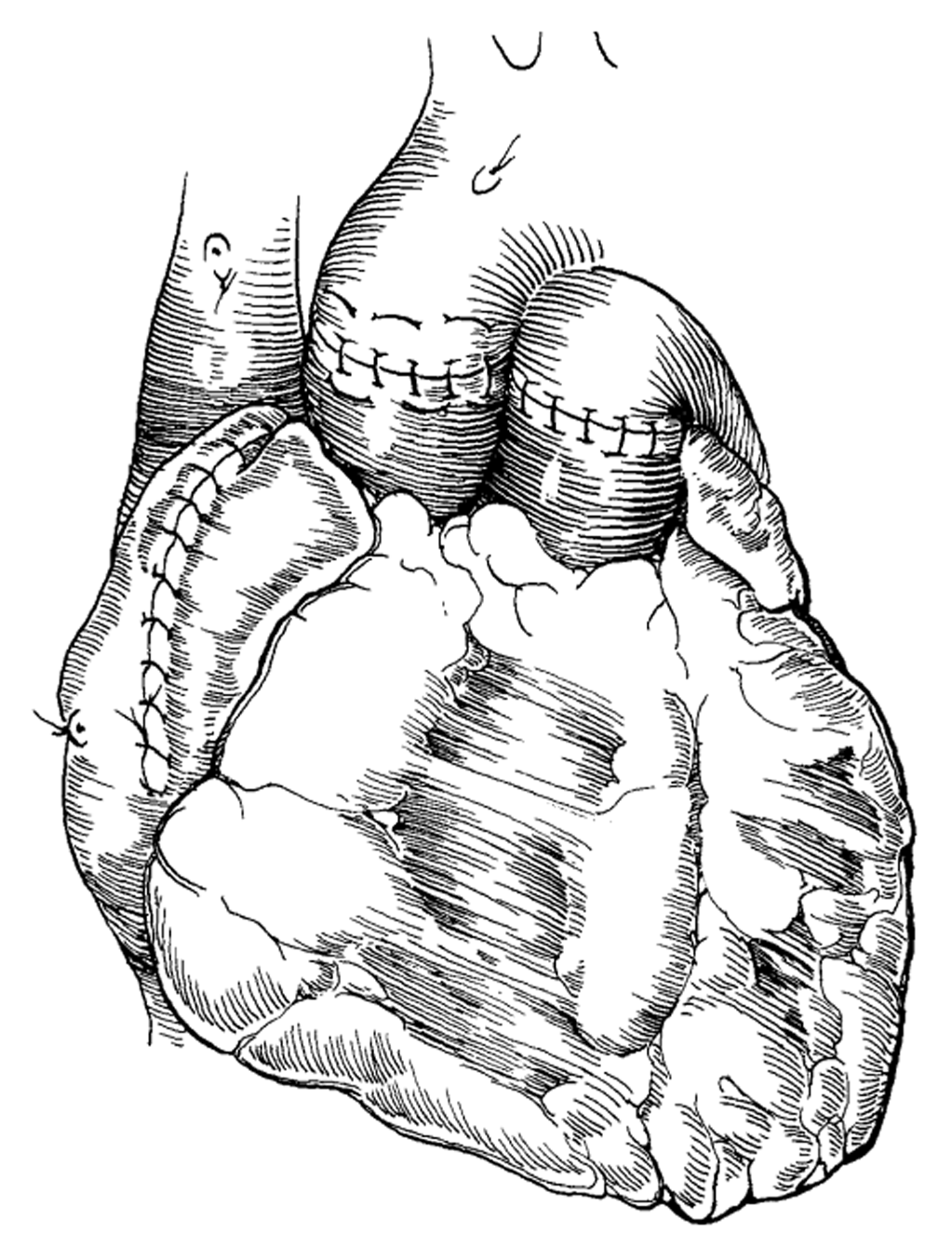
Figure 7
All the anastomoses are completed for the biatrial technique. Reprinted from Morton Bolman R. Oper Tech Thorac Cardiovasc Surg. 1999 May;4(2):98-113. ©1999, with permission from Elsevier.
Bicaval Implantation Technique
Hans Sievers and colleagues introduced the bicaval implantation technique into clinical practice in 1991. It was characterized by two arterial, one left atrial, and two caval anastomoses.6 The standard technique for orthotopic cardiac transplantation is done with large atrial anastomotic suture lines, which disrupts the anatomical integrity of the donor atria (Figure 8) and contributes electrophysiological disturbances and mechanical atrial dysfunction.7 In 1993, the description of the bicaval technique by Mazin Sarsam and colleagues had replaced the original biatrial technique. It was more anatomical and associated with less sinus node dysfunction, less atrial arrhythmias, and less tricuspid regurgitation.8
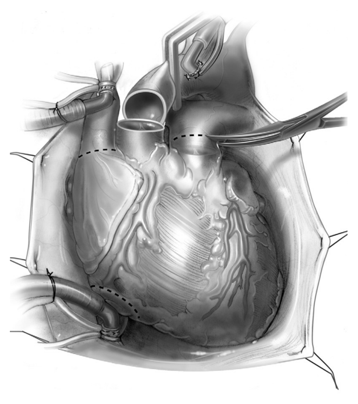
Figure 8
Donor cardiectomy used for bicaval implantation. Reprinted from John R, Liao K. Orthotopic Heart Transplantation. Oper Tech Thorac Cardiovasc Surg. 2010;15(2):138-146. ©2010, with permission from Elsevier.
Magdi Yacoub and his colleagues introduced the total heart transplant technique in 1989. It was purported to be a more anatomical surgical technique than that of bicaval implantation. It involved complete excision of the recipient’s right and left atria and direct anastomoses to the left pulmonary veins, right pulmonary veins, IVC and SVC.9 The rationale for this technique is to avoid nonphysiological geometry, which can lead to a higher incidence of mitral and tricuspid valve incompetence, rhythm disturbances, and the tendency of thrombus formation and septal aneurysm. However, it never gained popularity due to its complexity.
The standard recipient’s cardiectomy is done transecting the SVC and IVC in addition to the aorta and pulmonary artery transection (Figure 9).7
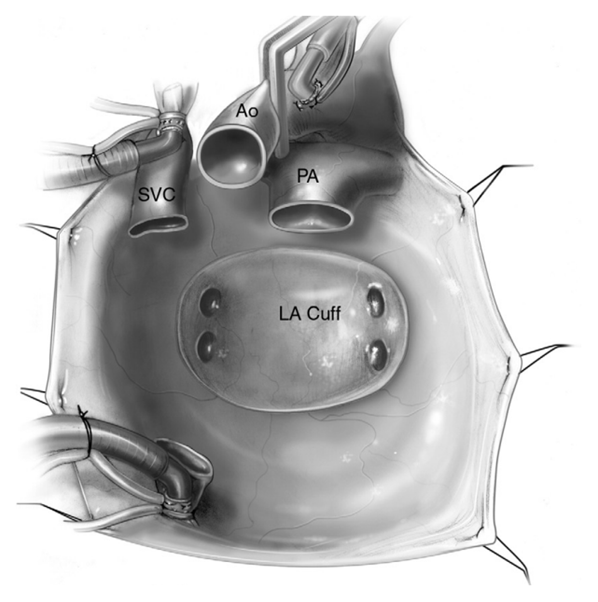
Figure 9
Recipient mediastinum after cardiectomy. Reprinted from John R, Liao K. Orthotopic Heart Transplantation. Oper Tech Thorac Cardiovasc Surg. 2010;15(2):138-146. ©2010, with permission from Elsevier. LA: left atrium; PA: pulmonary artery; Ao: aorta; SVC: superior vena cava
The left atrial anastomosis (Figure 10) is always performed by first using a long prolene suture starting at the left atrial cuff adjacent to the left superior pulmonary vein and passing it through the donor left atrial cuff adjacent to the left atrial appendage.7 The initial few sutures are completed with the donor heart positioned at the level of the sternal edge. The heart is subsequently lowered into the pericardial space. The posterior left atrial suture line is completed, and then the anterior suture line is facilitated by the assistant, who retracts the donor aorta and pulmonary artery to provide adequate exposure of the left atrium. To minimize the risk of left atrial thrombus formation, an everting suture technique should be used to facilitate approximation of the smooth endocardial surfaces of donor and recipient left atrial tissue.
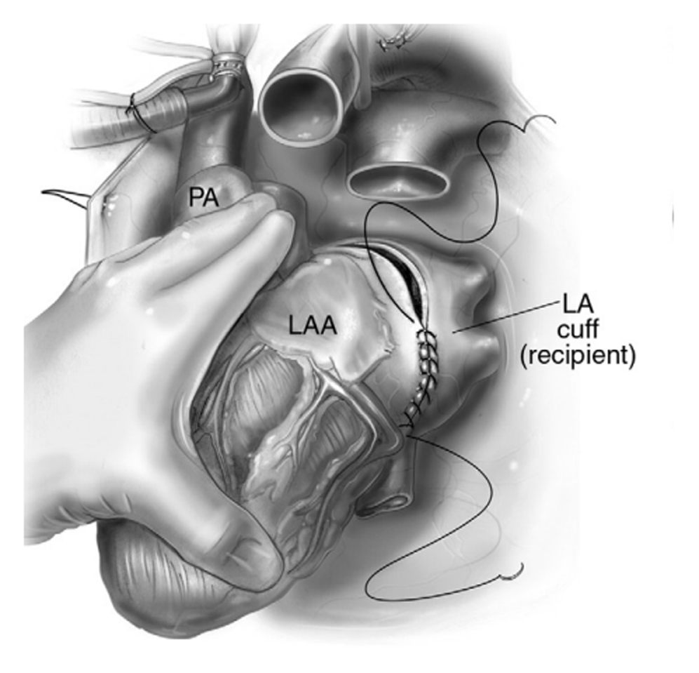
Figure 10
Bicaval technique using left atrial anastomosis. Reprinted from John R, Liao K. Orthotopic Heart Transplantation. Oper Tech Thorac Cardiovasc Surg. 2010;15(2):138-146. ©2010, with permission from Elsevier. LAA: left atrial appendage; LA: left atrium; PA: pulmonary artery
During all the anastomoses, a cold saline-soaked laparotomy pad can be placed on the heart. In addition, continuous carbon dioxide is running into the pericardial well.
Next, the inferior and superior vena caval anastomoses (Figure 11) are usually performed with prolene sutures.7 While performing the IVC anastomosis, care is taken to avoid deep sutures being placed in the region of the donor coronary sinus ostium to avoid potential injury. The donor right atrial appendage is medially oriented to facilitate proper orientation of the SVC anastomosis. This is important to avoid the SVC from being kinked or improperly aligned. Furthermore, close attention should be paid to avoid “purse-stringing” the SVC anastomosis to prevent inadvertent narrowing at the anastomotic level.
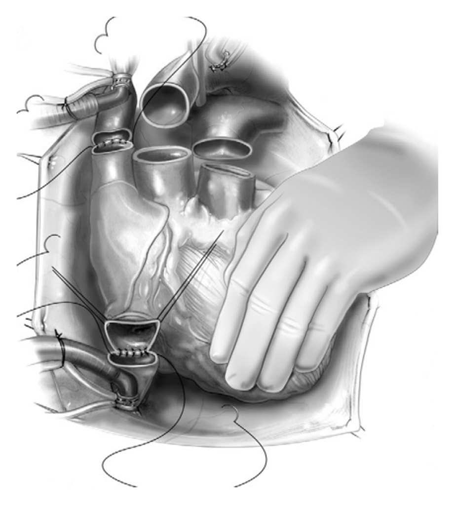
Figure 11
Inferior and superior vena cavae anastomosis used for bicaval technique. Reprinted from John R, Liao K. Orthotopic Heart Transplantation. Oper Tech Thorac Cardiovasc Surg. 2010;15(2):138-146. ©2010, with permission from Elsevier.
For pulmonary artery and aortic anastomosis (Figure 12), avoid excess length of the pulmonary artery.7 It is important to keep it short in order to avoid kinking at the level of the anastomosis, although there may be certain situations when more length of the pulmonary artery may be needed, such as congenital transplants. Leave raphe of the recipient’s pulmonary artery cusp commissures on the pulmonary artery to help orient the anastomosis in order to avoid twisting it. The anastomosis is performed with a 5-0 Prolene suture. Next, the aortic anastomosis is completed with a 4-0 or 5-0 Prolene suture (Figure 13).7
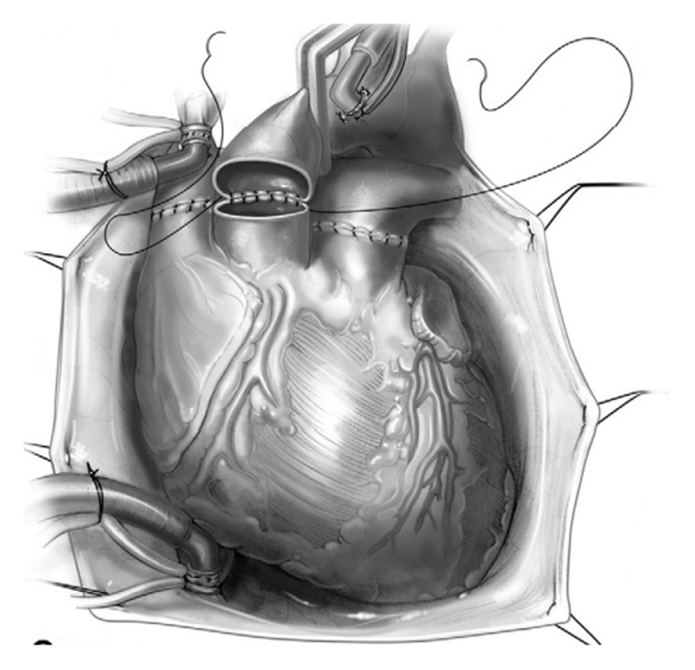
Figure 12
Aorta and pulmonary artery anastomosis used for bicaval technique. Reprinted from John R, Liao K. Orthotopic Heart Transplantation. Oper Tech Thorac Cardiovasc Surg. 2010;15(2):138-146. ©2010, with permission from Elsevier.
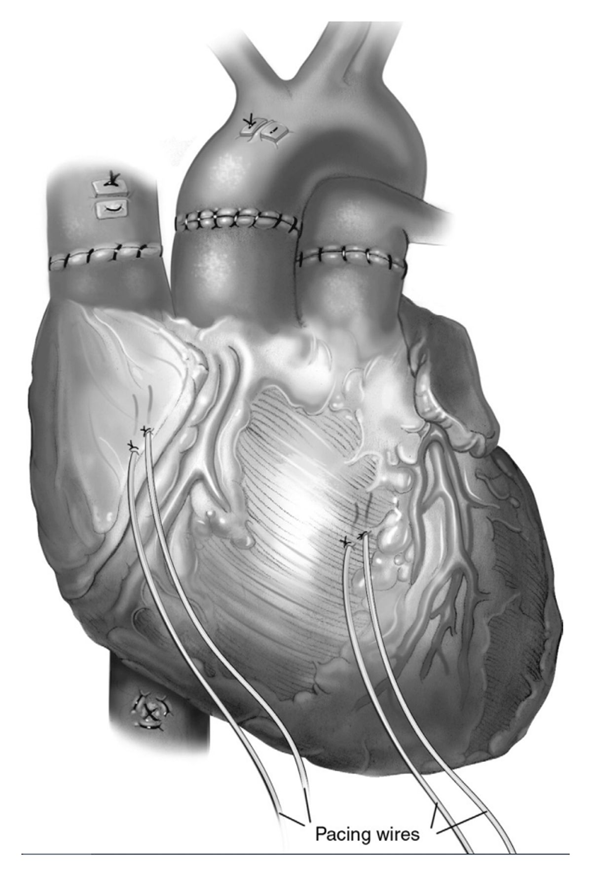
Figure 13
All the anastomosis are completed for the bicaval technique. Reprinted from John R, Liao K. Orthotopic Heart Transplantation. Oper Tech Thorac Cardiovasc Surg. 2010;15(2):138-146. ©2010, with permission from Elsevier.
Modified Bicaval Implantation Technique
The modified bicaval technique was first introduced in Japan in 2001 by Soichiro Kitamura and Takashi Kakuta, who left the posterior atrial bridge of tissue between the SCV and IVC intact.10 The advantages of this technique are to prevent retraction of the caval stumps that make the anastomosis difficult, to maintain the orientation of the caval stumps to prevent twisting of the anastomosis, and to allow for adjusting the sites where the caval anastomoses are done in case of extreme donor-recipient size mismatch, when the donor heart is much smaller than the recipient’s. With the presence of caval snares, the anatomical orientation can be lost, thus the anastomoses may be twisted or kinked. Furthermore, the caval end-to-end anastomosis in the original bicaval technique may become stenotic due to inadequate donor intercaval length in a size-mismatched donor, causing excessive tension on the suture line. By leaving a thin strip of the posterior wall of the right atrium as a bridge connecting the superior and inferior venae cavae, it is easy to adjust the amount of atrial excision to the donor heart size. The modified bicaval anastomosis technique allows for an adjustable caval anastomosis to compensate for the size mismatch. The modified bicaval technique may result in lower incidence of late caval anastomotic stricture because the anastomosis can be performed with no tension or kinking.11
Heterotopic Heart Transplantation Technique
After an historical series of experimental techniques and studies on animals, especially Demikhov’s early work, clinical heterotopic heart transplantation was introduced by Barnard and colleagues at the Groote Schuur Hospital, Cape Town, South Africa, in 1974.12 This was in response to a clinical need that was not met by orthotopic heart transplantation—namely, heart failure with pulmonary hypertension. Currently, heterotopic heart transplants are performed rarely, but they may be indicated in patients with irreversible fixed pulmonary hypertension or significant donor-recipient size mismatch when left ventricular assist devices are not available or contraindicated as a biological assist device.13,14
The donor heart is settled in the right thoracic cavity in the window created anterior to the right lung hilum, paying attention to avoid injury to the right phrenic nerve. The right-sided pleuro-pericardial flap is created. Cannulation strategies are similar to standard orthotopic heart transplantation. An incision is made into the Sondergaard groove of the recipient’s left atrium, similar to the standard left atriotomy incision. The recipient’s left atrium is sutured using a double-ended 4-0 polypropylene suture. The posterior aspect is completed first, then the anterior aspect.
The second anastomosis is the right atrial anastomosis. A 5-cm longitudinal incision is made into the lateral aspect of the recipient SVC and the right atrium, just anterior to the interatrial groove. The diamond shape anastomosis is made starting at the posterior wall and then finishing anterior, beginning 2 to 3 cm above the junction of the vena cava and extending 3 cm into the right atrium. The donor SVC and azygos veins are tied.
The third anastomosis connects the aortas. Proper length adjustment is done ventilating the lungs, which will push the recipient heart more medially. End-to-side anastomosis is done using a 4-0 Prolene suture. The last connection is the pulmonary arteries, using the proper size Dacron graft in an end-to-side fashion to the recipient’s main pulmonary artery.
Conclusion
Donor heart cardiectomy technique overall has not changed since Dr Shumway’s description. The surgical technique of orthotopic implantation has evolved considerably since 1967. In particular, the bicaval anastomosis technique has improved long-term survival related to better preservation of right atrial morphology. Nowadays mostly modified bicaval implantation techniques are used worldwide. Heterotopic heart transplantation has lost its importance since left ventricular assist device technology has received prominent acceptance.
Key Points
Since the first heart transplant was performed in 1967, the donor heart has been harvested using the biatrial technique for cardiac transplantation. Originally developed by Norman Shumway, this technique has been used in more than 20,000 patients.
The biatrial orthotopic heart transplantation technique has evolved considerably since 1967.
Introduced to clinical practice in 1991, the bicaval anastomosis implantation technique has improved long-term survival related to better preservation of right atrial morphology.
The modified bicaval implantation technique, now widely used, is thought to result in lower incidence of late caval anastomotic stricture because it can be performed without tension or kinking.
Competing Interests
The authors have no competing interests to declare.