
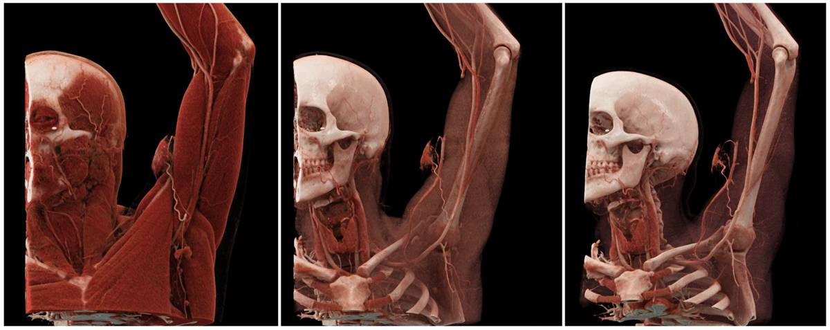
Figure 1
Three-dimensional cinematic reconstruction images. Left: The superficial layer, with just the skin and subcutaneous tissue removed, reveals the feeding artery originating from the proximal brachial artery. Middle: Increasing the opacity of the muscular layer reveals the malformation’s connection with the cephalic vein. Right: Further increasing the opacity permits better visualization of vascular structures.
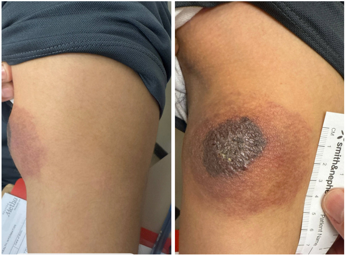
Figure 2
Protruding discolored mass located on the volar aspect of the upper arm shows signs of skin erosion.
Video 1
Axial slices of contrast-enhanced computed tomography angiography used for 3D cinematic reconstruction; see also at https://youtu.be/aEldgA6Az88.