
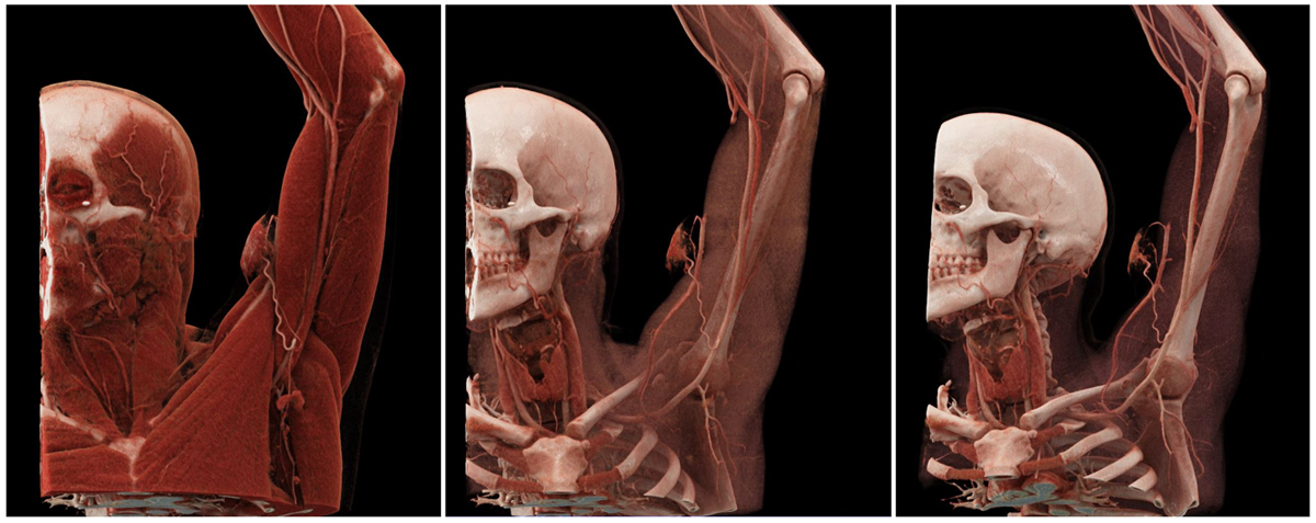
Figure 1
Three-dimensional cinematic reconstruction images. Left: The superficial layer, with just the skin and subcutaneous tissue removed, reveals the feeding artery originating from the proximal brachial artery. Middle: Increasing the opacity of the muscular layer reveals the malformation’s connection with the cephalic vein. Right: Further increasing the opacity permits better visualization of vascular structures.
Case Details
A 28-year-old male patient with no relevant medical history presented with recurrent bleeding from a mass located in the volar aspect of the proximal left upper arm. On physical examination, an approximately 5 × 5 cm protruding discolored lump was found, with no signs of active bleeding (Figure 2). It appeared superficial to the brachial fascia, making it amenable to open surgical resection. A thin-slice computed tomography (CT) angiography was performed to confirm the suspicion of vascular malformation, determine its nature (venous, arterial, or arteriovenous) and extent, and to understand its feeding and draining vessels towards planning an intervention (Video 1).
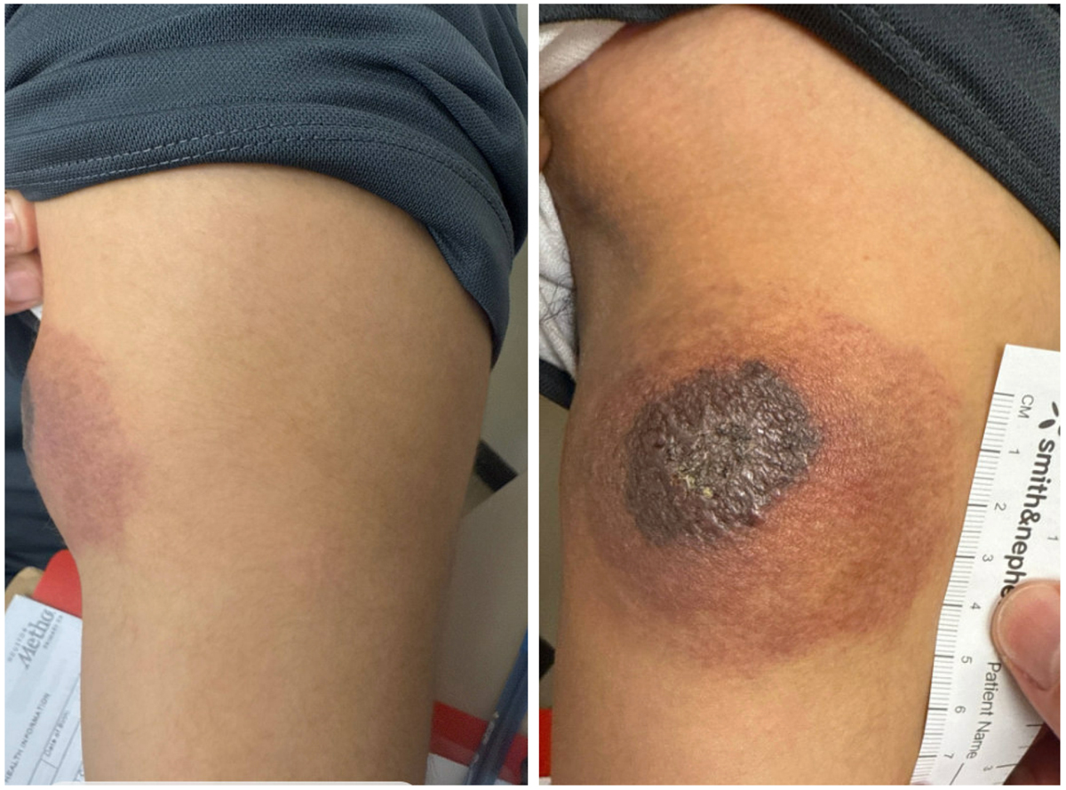
Figure 2
Protruding discolored mass located on the volar aspect of the upper arm shows signs of skin erosion.
Video 1
Axial slices of contrast-enhanced computed tomography angiography used for 3D cinematic reconstruction; see also at https://youtu.be/aEldgA6Az88.
A 3D cinematic-rendered CT image demonstrated a proximal branch of the brachial artery that was identified as the feeding vessel of the vascular malformation. Early enhancement of the cephalic vein proved the arteriovenous nature of the lesion. We performed 3D cinematic rendering using a commercially available software (Cinematic VRT, syngo.via VB50, Siemens Healthineers), which showed the feeding and draining vessels of the arteriovenous malformation and its relationship to the surrounding musculoskeletal structures (Figure 1).
In conclusion, 3D cinematic rendering facilitates the understanding of complex vascular pathologies; due to its intuitive nature, it can be a valuable tool for case planning and the education of medical professionals and patients alike. Of note, the quality of source images such as slice thickness (~1 mm) and optimal contrast enhancement also plays a crucial role in the generation of robust cinematic-rendered 3D images.
Competing Interests
Dr. Chinnadurai is a consultant for OccamLabs and Siemens Medical Solutions USA, Inc. Dr. Lumsden is a consultant for W. L. Gore & Associates, Siemens USA, and Boston Scientific and has ownership interest in Hatch Medical, Egg Medical, and BRIJ Medical. Dr. Lengyel has no competing interests to declare.