
Have a personal or library account? Click to login
Hypercoagulability and the A1298C MTHFR Mutation: Case Series of Unexplained Pulmonary Embolism Cover

Hypercoagulability and the A1298C MTHFR Mutation: Case Series of Unexplained Pulmonary Embolism

Open Access
|May 2025

Figures & Tables

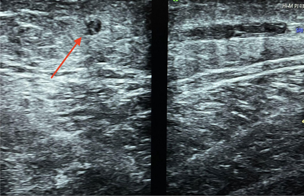
Figure 1

Echogenic thrombus in the great saphenous vein (red arrow).
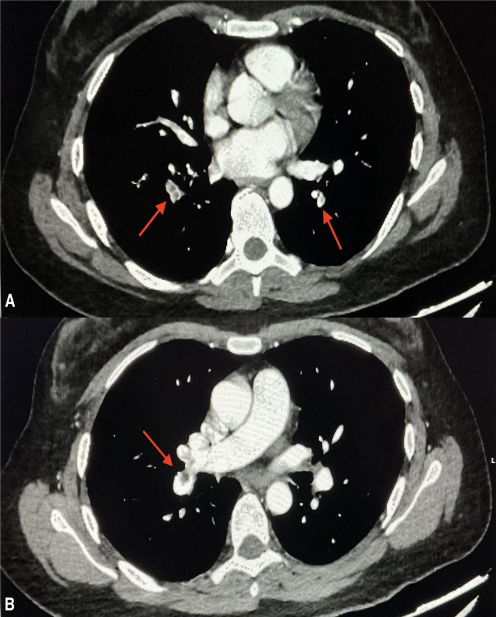
Figure 2

Coronary computed tomography pulmonary angiography showing segmental filling defects in the pulmonary artery branches of both upper and lower lobes: (A) filling defects in the segmental branches of the pulmonary artery are suggestive of thrombus (red arrows) and (B) eccentric filling defect in the interlobar artery (red arrow).
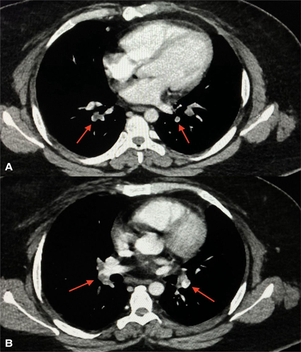
Figure 3

Contrast-enhanced computed tomography chest hypodense filling defects in the upper and lower lobar pulmonary artery branches bilaterally: (A) hypodense filling defects in the bilateral pulmonary artery branches (red arrows) and (B) extension of thrombus in the right and left pulmonary artery branches (red arrows).

DOI: https://doi.org/10.14797/mdcvj.1565 | Journal eISSN: 1947-6108
Language: English
Page range: 57 - 62
Submitted on: Jan 11, 2025
Accepted on: Mar 27, 2025
Published on: May 30, 2025
Published by: Houston Methodist DeBakey Heart & Vascular Center
In partnership with: Paradigm Publishing Services

© 2025 Akshat Sahai, Vaibhav Sharma, Prapti Mishra, Avinash Siddanoor, Abhishek Bhatia, Deepak Gargi Pande, published by Houston Methodist DeBakey Heart & Vascular Center
This work is licensed under the Creative Commons Attribution-NonCommercial 4.0 License.