
Introduction
Methylenetetrahydrofolate reductase (MTHFR) is a key enzyme in homocysteine metabolism. It converts to methionine via the folate pathway. Mutations in the MTHFR gene, such as A1298C and C677T polymorphisms, have been associated with reduced enzymatic activity, leading to hyperhomocysteinemia—a recognized risk factor for venous thromboembolism.1,2,3 While the C677T mutation has been widely studied and linked to thrombotic events, the clinical significance of the A1298C polymorphism remains controversial, with conflicting evidence in the literature regarding its contribution to hypercoagulable states.4
Venous thromboembolism (VTE), encompassing deep vein thrombosis (DVT) and pulmonary embolism (PE), is a potentially life-threatening condition with multifactorial etiologies. This study involved collecting and analyzing comprehensive clinical, laboratory, and radiological data for two patients with PE who were found to have the A1298C MTHFR gene mutation.
Case 1 involved a 42-year-old female with a homozygous A1298C mutation who presented with complaints of DVT complicated by bilateral PE and demonstrated moderately increased homocysteine levels. Case 2 involved a 34-year-old woman with a heterozygous A1298C mutation who presented with isolated PE and markedly elevated homocysteine levels. Both patients are free from traditional VTE risk factors and demonstrated negative results for other hereditary and acquired thrombophilias. Anticoagulation therapy resulted in complete resolution of symptoms with no recurrence at follow-up.
Patient A presented with pain in the right lower limb and shortness of breath. Venous Doppler showed thrombosis of the great saphenous vein and bilateral pulmonary embolism on computed tomography (CT) pulmonary angiography (CTPA). Patient B presented with fever, shortness of breath, and painful swelling in both lower limbs for the past week. Contrast-enhanced computed tomography (CECT) revealed multiple thrombi in bilateral upper and lower lobar pulmonary artery branches. Both patients were managed medically and were discharged with no complications. At follow-up, they continue to be asymptomatic. Both patients had no identifiable secondary causes of hypercoagulability, strengthening the likelihood of a causal association with the genetic mutation. Through these cases we highlight the variability in clinical presentations and underline the potential role of genetic testing in identifying underlying causes of unexplained VTE.
Case Presentation
Patient A
A 42-year-old female with no significant past medical history presented to the emergency department with complaints of dyspnea and right lower extremity pain for the duration of 5 days. The lower extremity pain was initially localized to the right thigh and subsequently progressed to involve the right knee and calf region. The patient denied any recent trauma, prolonged bed rest, surgical procedures, or use of hormone replacement therapy or oral contraceptive pills. Family history was not contributory to thrombotic disorders or recurrent pregnancy losses.
On physical examination, the patient’s vitals were within normal limits. The right lower extremity was significantly tender and erythematous with a positive Homan’s sign. The cardiopulmonary examination was unremarkable, with clear breath sounds bilaterally and regular heart rhythm without any murmurs.
These findings were confirmed by diagnostic imaging. Venous Doppler ultrasonography demonstrated an echogenic thrombus in the right tibioperoneal trunk and the great saphenous vein shown in Figure 1. CTPA confirmed bilateral pulmonary embolism, with filling defects noted in both upper and lower lobar branches of the pulmonary artery, as illustrated in Figure 2. Electrocardiogram (ECG) and two-dimensional echocardiography (2D-ECHO) were unremarkable, with no evidence of right ventricular strain.
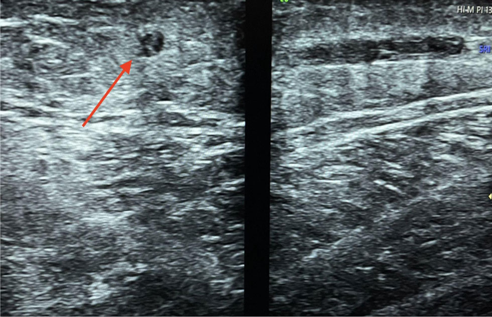
Figure 1
Echogenic thrombus in the great saphenous vein (red arrow).
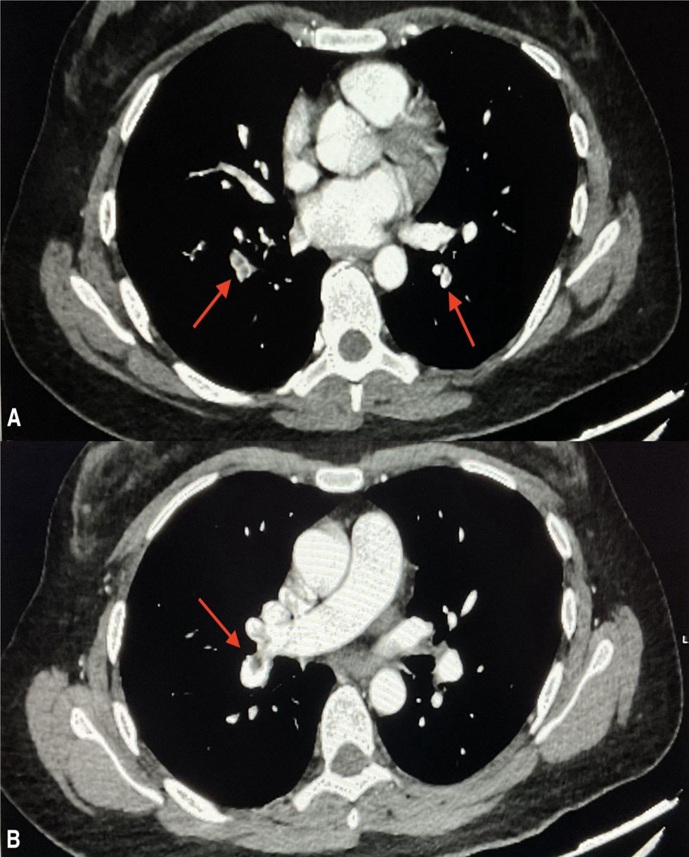
Figure 2
Coronary computed tomography pulmonary angiography showing segmental filling defects in the pulmonary artery branches of both upper and lower lobes: (A) filling defects in the segmental branches of the pulmonary artery are suggestive of thrombus (red arrows) and (B) eccentric filling defect in the interlobar artery (red arrow).
Laboratory Tests
The initial laboratory studies were suggestive of microcytic hypochromic anemia with hemoglobin values at 8.3 g/dL (normal range 12.5–15.5 g/dL) and serum iron at 38 mcg/dL (normal range 60–170 mcg/dL). Basic laboratory tests, including complete blood count (CBC), comprehensive metabolic panel (CMP), liver function tests (LFT), and inflammatory markers, were normal. The coagulation profile revealed a D-dimer level of 804 ng/mL (normal range ≤ 300 ng/mL). Cultures of blood, urine, and sputum were sterile. A comprehensive study for thrombophilia was also done, which returned negative for Factor V Leiden mutation, lupus anticoagulant, and antiphospholipid antibodies (APLA).
Antithrombin III activity was 67% (reference range: 10–150%), and the levels of protein C and S were within the normal range. Antinuclear antibody (ANA) and antineutrophil cytoplasmic antibody (ANCA) were negative. Homocysteine, however, was elevated at 30.7 μmol/L (reference range of 4.44–13.56 μmol/L). Genetic analysis later revealed that the patient was homozygous for the mutation A1298C in the MTHFR gene, whereas the C677T variant was negative.
Management
The patient was initiated on anticoagulation therapy with subcutaneous enoxaparin and later transitioned to oral apixaban. The patient continued to improve symptomatically and was discharged from the hospital on day 5 after making proper follow-up arrangements. She remained asymptomatic at 2 weeks post-admission without any recurrence of thrombosis.
Patient B
A 34-year-old female with no known comorbidities presented to the emergency department with a 4-day history of fever and progressive dyspnea. She denied any history of cough, recent travel, trauma, or prolonged immobilization. There was no history of oral contraceptive or hormone therapy use, recent surgical procedures, or recurrent pregnancy losses. No significant family history of thrombotic disorders was reported.
Physical examination revealed a temperature of 101.4° F, tachycardia (102/min), and tachypnea (24/min). Blood pressure and oxygen saturation were within normal limits. The cardiopulmonary examination revealed clear breath sounds on both sides with no adventitious sounds.
Diagnostic imaging studies revealed significant results. Though the venous Doppler ultrasonography of the lower extremities was negative for DVT, the CECT of the chest revealed multiple hypodense filling defects in the bilateral upper and lower lobar pulmonary artery branches, consistent with PE (Figure 3). ECG and 2D-ECHO were unremarkable, with no evidence of right ventricular strain.
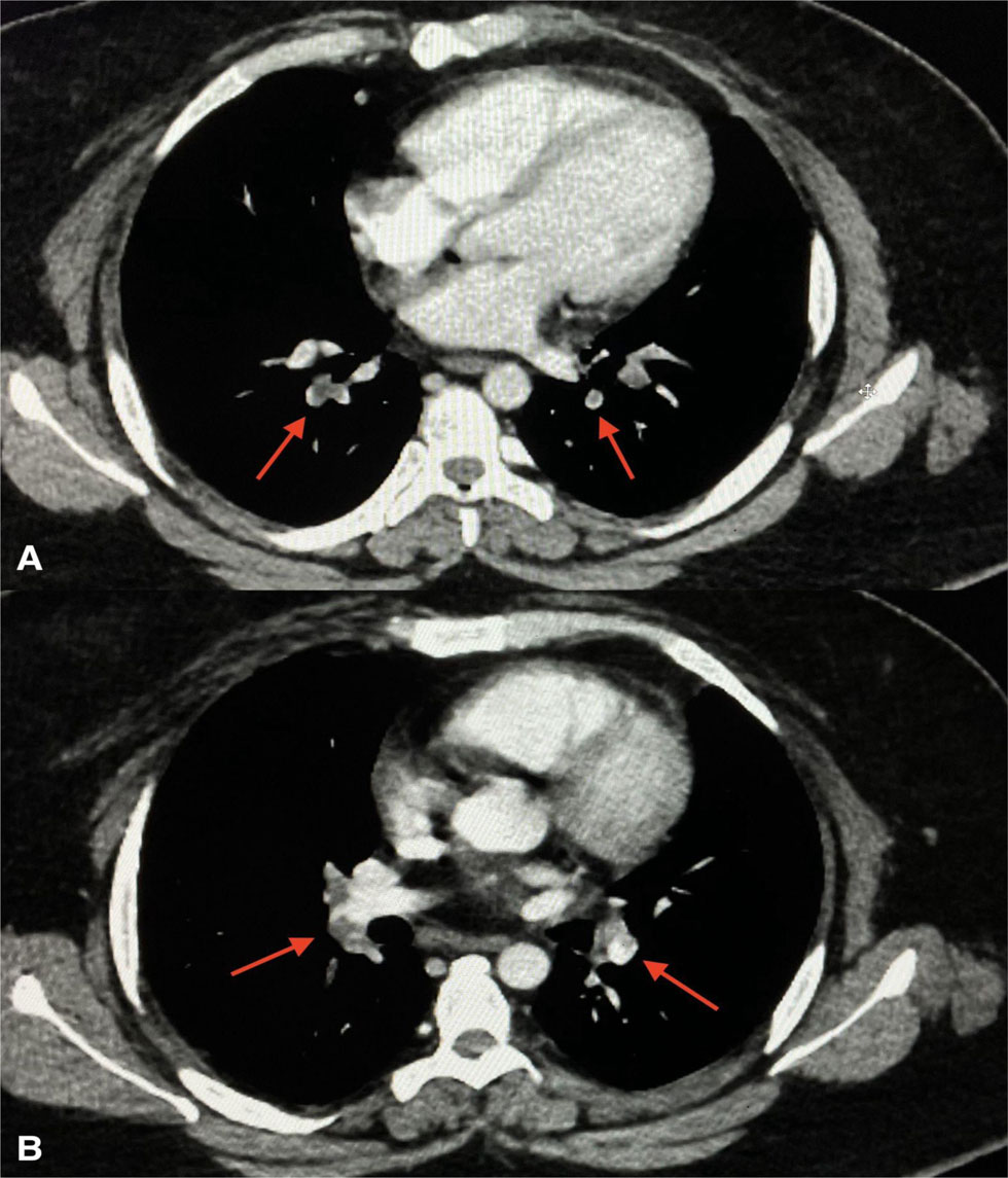
Figure 3
Contrast-enhanced computed tomography chest hypodense filling defects in the upper and lower lobar pulmonary artery branches bilaterally: (A) hypodense filling defects in the bilateral pulmonary artery branches (red arrows) and (B) extension of thrombus in the right and left pulmonary artery branches (red arrows).
Laboratory Tests
Laboratory evaluation demonstrated normocytic anemia with hemoglobin of 9.2 g/dL (reference range: 14–18 g/dL) and serum iron of 41 mcg/dL (reference range: 60–170 mcg/dL). Basic laboratory parameters, including CBC, CMP, and inflammatory markers, were within normal limits, and blood, urine, and sputum cultures were sterile. Coagulation studies highlighted elevated D-dimer levels at 1008 ng/mL (reference range: ≤ 300 ng/mL). A comprehensive thrombophilia workup excluded other hypercoagulable states, including Factor V Leiden mutation, antiphospholipid syndrome, and deficiencies in antithrombin III, protein C, and protein S. ANA and ANCA were negative.
The most significant finding was an elevated homocysteine level at 182 μmol/L (normal range: 4.44–13.56 μmol/L). Genetic studies were positive for a heterozygous A1298C mutation in the MTHFR gene but negative for the C677T variant. Cardiac biomarkers were elevated NT-proBNP at 1457 pg/mL (reference range < 125 pg/mL) and troponin I at 16.2 pg/mL (reference range 0–40 pg/ml).
Management
The patient was initiated on anticoagulation therapy with subcutaneous enoxaparin and later switched over to oral apixaban. She showed clinical improvement with resolution of fever and dyspnea and was discharged on day 7 of hospitalization with proper follow-up arrangements. At 2 weeks follow-up, the patient remained asymptomatic with no signs of recurrent thromboembolism.
Comparative Overview
This series of cases offers two female patients with PE linked to the A1298C mutation of the MTHFR gene, where obvious changes appear in clinical presentation, laboratory analysis, and genetic characters. Some of the key distinctions and similarities are highlighted as follows:
Clinical Presentation and Demographics: Patients were two female premenopausal patients. Their ages of 42 and 34 years did not have an historical risk factor to cause thromboembolism.
Presenting Complaints: Patient A had classical DVT symptomatology that evolved into PE, while Patient B had systemic symptoms (pyrexia) and isolated PE without radiological evidence of DVT.
Imaging Findings: Patient A had DVT and bilateral PE with a thrombus in the tibioperoneal trunk and great saphenous vein. Patient B only had PEs without imaging evidence of DVT but rather may be an in-situ thrombosis in the pulmonary artery.
Coagulation Studies: Both had increased D-dimer levels (804 ng/mL and 1008 ng/mL). They also had elevated homocysteine levels, which significantly impacted hypercoagulability. Neither patient had other inherited or acquired thrombophilias.
Genetic Findings: Patient A had a homozygous A1298C MTHFR mutation, whereas Patient B had a heterozygous mutation.
Management and Outcomes: Both patients underwent standardized anticoagulation protocols, with initial treatment with enoxaparin followed by a transition to apixaban. Neither patient required thrombolysis or mechanical intervention. Both patients demonstrated optimum responses to anticoagulation. No complications were reported during the acute phase. Two-week follow-up resulted in a complete resolution of symptoms, and no recurrent thrombotic events were noted.
Discussion
This case series highlights two patients presenting with PE who were found to have the A1298C mutation in the MTHFR gene. One patient was homozygous for the mutation, and the other was heterozygous, yet both demonstrated hyperhomocysteinemia, suggesting a potential role of this genetic variation in hypercoagulability and thromboembolic events. While the clinical significance of MTHFR mutations, particularly A1298C, remains debated, these cases contribute to the growing discourse on its potential association with VTE.1
The MTHFR enzyme plays a pivotal role in converting homocysteine to methionine via the folate pathway. Mutations such as A1298C and C677T are known to reduce enzymatic activity, potentially leading to hyperhomocysteinemia, which is a recognized prothrombotic condition. Elevated homocysteine levels can exacerbate thrombogenesis through increased tissue factor expression, reduced plasmin generation, heightened platelet reactivity, and increased oxidative stress.1 While the C677T mutation has been extensively studied and consistently associated with thrombotic events, the role of the A1298C polymorphism in thromboembolism remains less clear, with conflicting findings in the literature.4
The A1298C mutation is typically associated with a lesser impact on homocysteine levels than C677T. However, in homozygous states, it can still lead to significant hyperhomocysteinemia, as seen in Patient A.2,3 Intriguingly, Patient B, who was heterozygous for the A1298C mutation, also exhibited elevated homocysteine levels, suggesting that other genetic, metabolic, or environmental factors may have contributed to hypercoagulability in this case.5 Emerging evidence indicates that factors such as deficiencies in vitamins B6, B12, and folate, chronic inflammation, renal dysfunction, or coexisting polymorphisms can modulate the impact of MTHFR mutations on homocysteine metabolism and thrombotic risk.6,7
The clinical presentation in these cases underscores the heterogeneity of VTE. Patient A presented with pain and swelling in the lower limb due to DVT and subsequently developed bilateral PE, confirmed by CTPA. In contrast, Patient B presented with fever, shortness of breath, and bilateral limb pain, with CECT revealing multiple pulmonary arterial thrombi without evidence of DVT on the venous Doppler. Comprehensive evaluations excluded other common causes of hypercoagulability, including Factor V Leiden mutation, antiphospholipid syndrome, and deficiencies of antithrombin III, protein C, and protein S. The absence of these factors strengthens the association between the A1298C mutation and their thrombotic events.
Both patients were successfully managed with anticoagulation therapy, the cornerstone of VTE treatment.8 In addition to anticoagulation, addressing elevated homocysteine levels is critical in patients with MTHFR mutations. Supplementation with folic acid, vitamin B6, and vitamin B12 has been shown to lower homocysteine levels and may reduce the risk of recurrence.9 However, the efficacy of homocysteine-lowering therapy in preventing thrombotic events, particularly in the context of MTHFR polymorphisms, remains a subject of ongoing investigation.
Routine genetic screening for MTHFR mutations in patients with VTE is not currently recommended, as these polymorphisms are common in the general population and have variable clinical significance.10 Nevertheless, targeted testing may be warranted in cases of unexplained thrombosis, especially when other causes of hypercoagulability have been ruled out. Considering the patient’s genetic profile, family history, and overall risk factors, a personalized approach is essential for optimizing management strategies.11
This case series raises important questions about the thrombotic risk associated with the A1298C mutation, particularly in heterozygous states. It also highlights the potential utility of testing for MTHFR mutations in patients with unexplained VTE. Large-scale prospective studies are needed to better understand the interaction between genetic, nutritional, and environmental factors in modulating thrombotic risk. Research could help refine screening, prevention, and management guidelines for patients with MTHFR mutations.
Conclusion
In conclusion, while the role of the A1298C mutation in thromboembolism remains controversial, these cases suggest it may contribute to hypercoagulable states in elevated homocysteine levels. This underscores the importance of a multidisciplinary approach in evaluating and managing patients with unexplained VTE. Further studies are warranted to elucidate the significance of this genetic variation and its role in the risk stratifications for VTE.
Key Points
The A1298C MTHFR mutation may contribute to a hypercoagulable state, especially in unexplained thrombosis cases. Selective genetic testing and personalized management strategies, including anticoagulation and homocysteine-lowering therapy, can optimize care.
Although routine screening for MTHFR mutations is not recommended, targeted testing and a multidisciplinary approach may benefit patients with recurrent or unexplained thrombotic events, improving risk stratification and treatment strategies.
Large-scale studies are needed to understand better the clinical relevance of the A1298C mutation and its interaction with genetic, environmental, and nutritional factors as well as to guide evidence-based guidelines for screening and management.
Competing Interests
The authors have no competing interests to declare.