
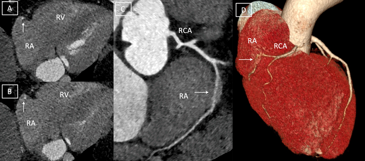
Figure 1
Axial computed tomography image (A, B) depicting intra-atrial course of right coronary artery (RCA, depicted by white arrow). Curvilinear multiplanar image (C) and volume-rendered image (D) demonstrate proximal RCA surrounded by epicardial fat, intra-atrial course of RCA (white arrow) for a length of 3 cm and then normal course of rest of RCA. RV: right ventricle; RA: right atrium