
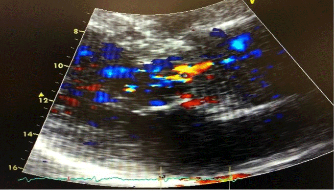
Figure 1
Pre-COVID-19 transthoracic echocardiogram showing mild aortic regurgitation.
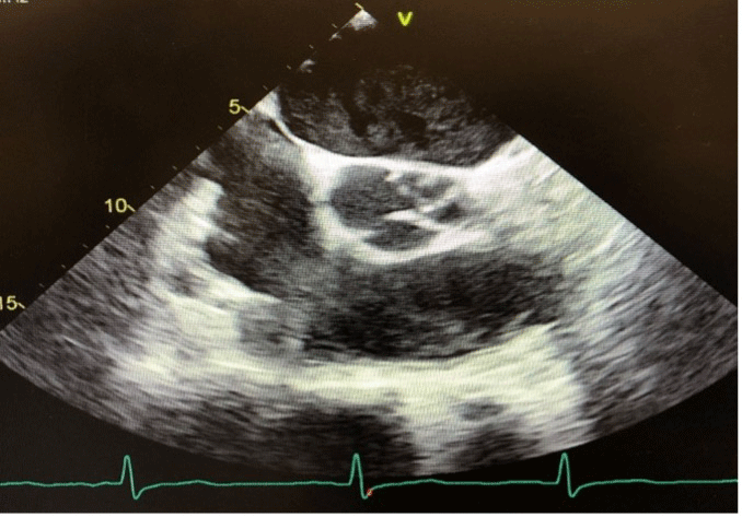
Figure 2
Post-COVID-19 transthoracic echocardiogram showing prolapse of the aortic valve leaflets during diastole.
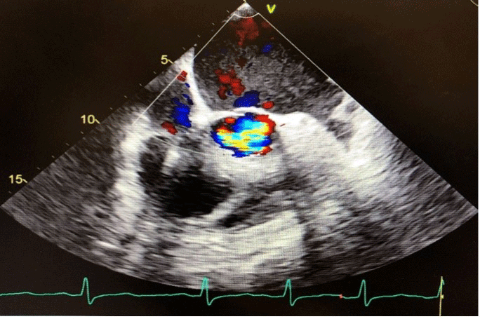
Figure 3
Post-COVID-19 transesophageal echocardiogram showing severe aortic regurgitation.
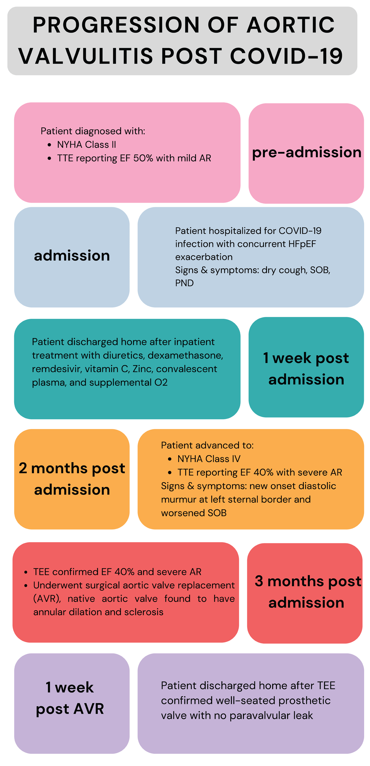
Figure 4
Progression of symptoms, workup, and management in patient with aortic valvulitis in the setting of COVID-19.
Table 1
Timeline of transthoracic echocardiogram findings in patient prior, during, and post COVID-19 infection. TTE: transesophageal echocardiogram; AV: aortic valve; LVEF: left ventricle ejection fraction; LV: left ventricle; AR: aortic regurgitation
| DATE OF STUDY | TTE 4/14/2019 | TTE 7/21/2020 | TTE 11/11/2020 *TIME OF COVID-19 INFECTION* | TTE 1/12/2021 | TEE 1/27/2021 *INTRA-OPERATIVE, DURING AV REPLACEMENT* | TTE 1/19/2022 | TTE 1/26/2023 | TTE 1/24/2024 | TTE 3/3/2025 |
|---|---|---|---|---|---|---|---|---|---|
| LVEF | 55 - 60% | 55 - 60% | 45 - 50% | 45% | 40 - 45% | 55% | 53.1% | 50% | 50 - 55% |
| LV diastolic diameter | 5.97 cm | 5.66 cm | 7.0 cm | 6.79 cm | 5.11 cm | 5.28 cm | 5.64 cm | 4.99 cm | |
| LV systolic diameter | 4.4 cm | 3.83 cm | 3.4 cm | 5.02 cm | 3.43 cm | 3.83 cm | 3.82 cm | 3.65 cm | |
| Left ventricle | Grade II diastolic dysfunction, normal LV systolic function | Normal diastolic and systolic LV function | Low-normal systolic LV function | Mild-moderate LV dilation, mild-moderate decrease in LV function | Diffuse hypokinesis, reduced LV systolic function | Grade I diastolic dysfunction, normal LV systolic function | Normal LV systolic and diastolic function | Grade I diastolic dysfunction, normal LV systolic function | |
| Aortic valve | Mild AR | Moderate AR | Moderate AR | Moderate-severe AR | Severe AR | Bioprosthetic AV, trace AR | Bioprosthetic valve, mild AR | Bioprosthetic valve, no valvular or peri-valvular AR | Bioprosthetic valve, mild AR |