
Introduction
In late 2019, a novel coronavirus caused by severe acute respiratory syndrome coronavirus 2 (SARS-CoV-2) was identified as the source of a viral illness spreading throughout Wuhan, China. The virus quickly disseminated throughout China and the rest of the world, erupting in a global pandemic. By March of 2021, there were approximately 120 million confirmed cases that resulted in more than 2.5 million deaths worldwide.1 The most common presenting symptoms of COVID-19 at the time were fever, cough, fatigue, myalgias, and headache, with pneumonia being the most common severe manifestation.2,3 Additionally, several complications of COVID-19 were later reported, most commonly respiratory failure, sepsis, myocardial injury, and heart failure.4 A recent 2023 study noted a particularly high rate of cardiovascular complications in COVID-19 patients, estimating the incidence of acute heart failure and myocardial injury in the setting of COVID-19 at 22% to 33% and 31%, respectively.5 These complications also include myocarditis, acute myocardial infarction, cardiomyopathy, cardiogenic shock, arrhythmias, cardiac arrest, and venous thromboembolic events.6
The exact mechanism for the predilection of COVID-19 in the human heart remains unknown. However, it has been proposed that the highly expressed angiotensin converting enzyme-2 (ACE2), which serves as the host cellular receptor for virus entry in the cardiovascular tissue, may increase the susceptibility of this system to complications of COVID-19.7 More specifically, ACE2 has been detected in cardiac valves, particularly the aortic valve, and proposed as a mediator in valvular integrity.8 Thus, it is reasonable to consider valvular disease as another cardiovascular complication associated with COVID-19. Here, we present a patient who developed severe aortic regurgitation (AR) over the course of 2 months following infection with COVID-19, with no other plausible etiologies identified. This is a critical contribution to the current literature as there are no other reported cases of COVID-19-related AR.
Case Report
We present a 69-year-old Caucasian male who came to the Emergency Department complaining of a dry cough, worsening shortness of breath, and paroxysmal nocturnal dyspnea after having been recently discharged from another hospital for an exacerbation of his heart failure with preserved ejection fraction (HFpEF). The patient provided a past medical history significant for atrial fibrillation anticoagulated on rivaroxaban, HFpEF, chronic kidney disease, and polycythemia vera. Prior to this acute illness, the patient’s activity level was classified as New York Heart Association (NYHA) class II with his most recent echocardiogram revealing an ejection fraction (EF) of 50%, mild AR, an LV diastolic diameter of 7 cm and systolic diameter of 3.4 cm (Figure 1). During this admission, the patient was diagnosed with COVID-19 as well as persistent exacerbation of his HFpEF. Management included diuretic therapy and guideline-recommended treatment for COVID-19 with dexamethasone, remdesivir, vitamin C, zinc, and a single unit of convalescent plasma. He briefly required supplemental oxygen with 2 liters nasal cannula. Ultimately, the patient improved and was discharged 1 week later.
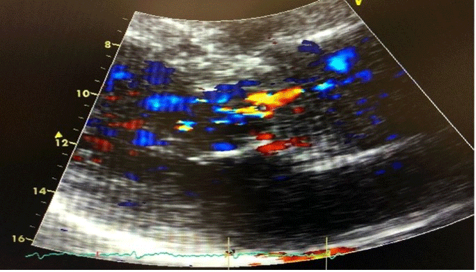
Figure 1
Pre-COVID-19 transthoracic echocardiogram showing mild aortic regurgitation.
Over the next 2 months, the patient developed progressively worsening shortness of breath with his activity level now at NYHA class IV. He was also noted to have a new-onset diastolic murmur heard best at the left sternal border on physical examination. In January of 2021, a follow-up transthoracic echocardiogram was ordered, which revealed an EF of 40%, severe AR, and an LV diastolic diameter of 7.52 cm and systolic diameter of 3.3 cm. Cardiac magnetic resonance imaging and computed tomography were not performed as they were unavailable at our institution and the patient presented in a decompensated state requiring expedited intervention.
The patient was subsequently referred to a cardiothoracic surgeon to evaluate for aortic valve replacement. In February of 2021, a transesophageal echocardiogram (TEE) was performed, which confirmed the diagnosis of severe AR with reduced EF at 40% (Figures 2, 3). Additionally, he underwent pre-procedural cardiac catheterization, which revealed mild, nonobstructive, single-vessel coronary artery disease. Ultimately, the patient received a surgical aortic valve replacement. During the procedure, the aortic valve was found to be a trileaflet valve with annular dilatation, and the surgical pathology report revealed myxoid degeneration; subsequently, this was replaced with a pericardial tissue heart valve. Intraoperative TEE following valve replacement revealed a well-seated prosthetic valve without evidence of paravalvular leak or valvular insufficiency. The postoperative course was without complication, and the patient was discharged from the hospital 6 days later.
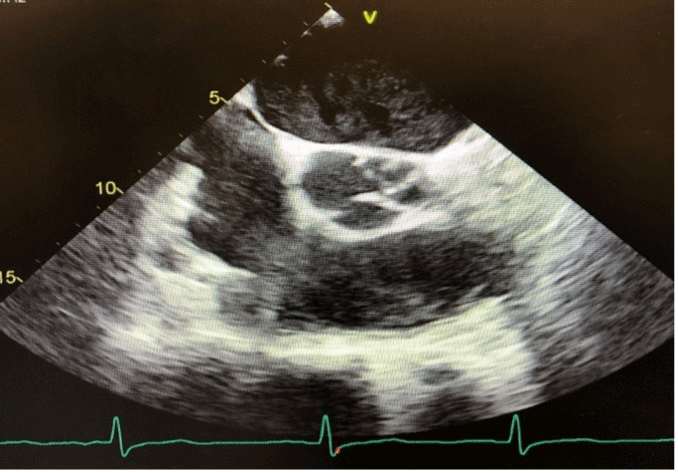
Figure 2
Post-COVID-19 transthoracic echocardiogram showing prolapse of the aortic valve leaflets during diastole.
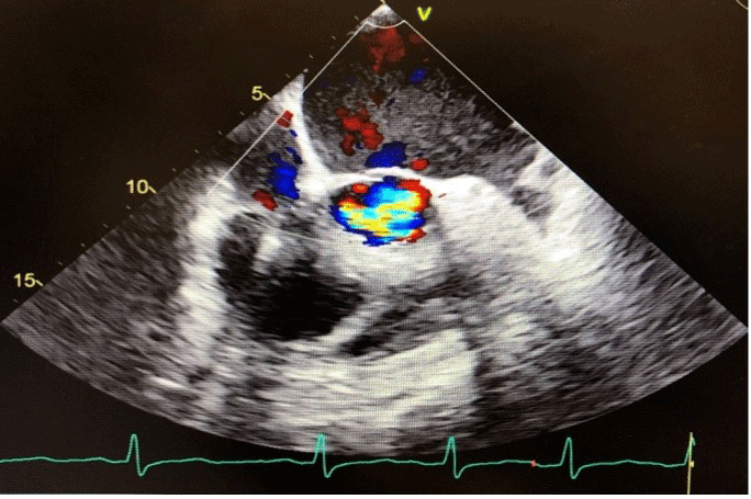
Figure 3
Post-COVID-19 transesophageal echocardiogram showing severe aortic regurgitation.
Discussion
In contrast to chronic AR, acute AR does not allow for compensatory dilatation of the left ventricle to accommodate the volume overload state caused by the regurgitant valve. There is an acute increase in left ventricular diastolic pressures, a subsequent fall in forward cardiac output, and heart failure symptomatology quickly ensues, as seen in this patient.9,10 Specifically, our patient experienced a 15% to 20% reduction in EF, considerable reduction in LV systolic function, and progression of AR from mild to severe (Figure 4, Table 1). The etiologies of acute native aortic valve regurgitation are limited and most commonly include endocarditis and aortic dissection.11 However, given the patient’s lack of autoimmune disease history and negative blood culture results, the likely cause of acute AR in this case favored his recent COVID-19 infection. His pathology report revealed slightly calcified aortic valvular tissue with myxoid degeneration. The chordae tendineae were slightly elongated with no apparent vegetations. Although calcific valve disease and myxomatous degeneration are among the most common causes of chronic AR, these findings do not account for the acute decline in valvular disease and hemodynamic dysfunction seen in this patient.12
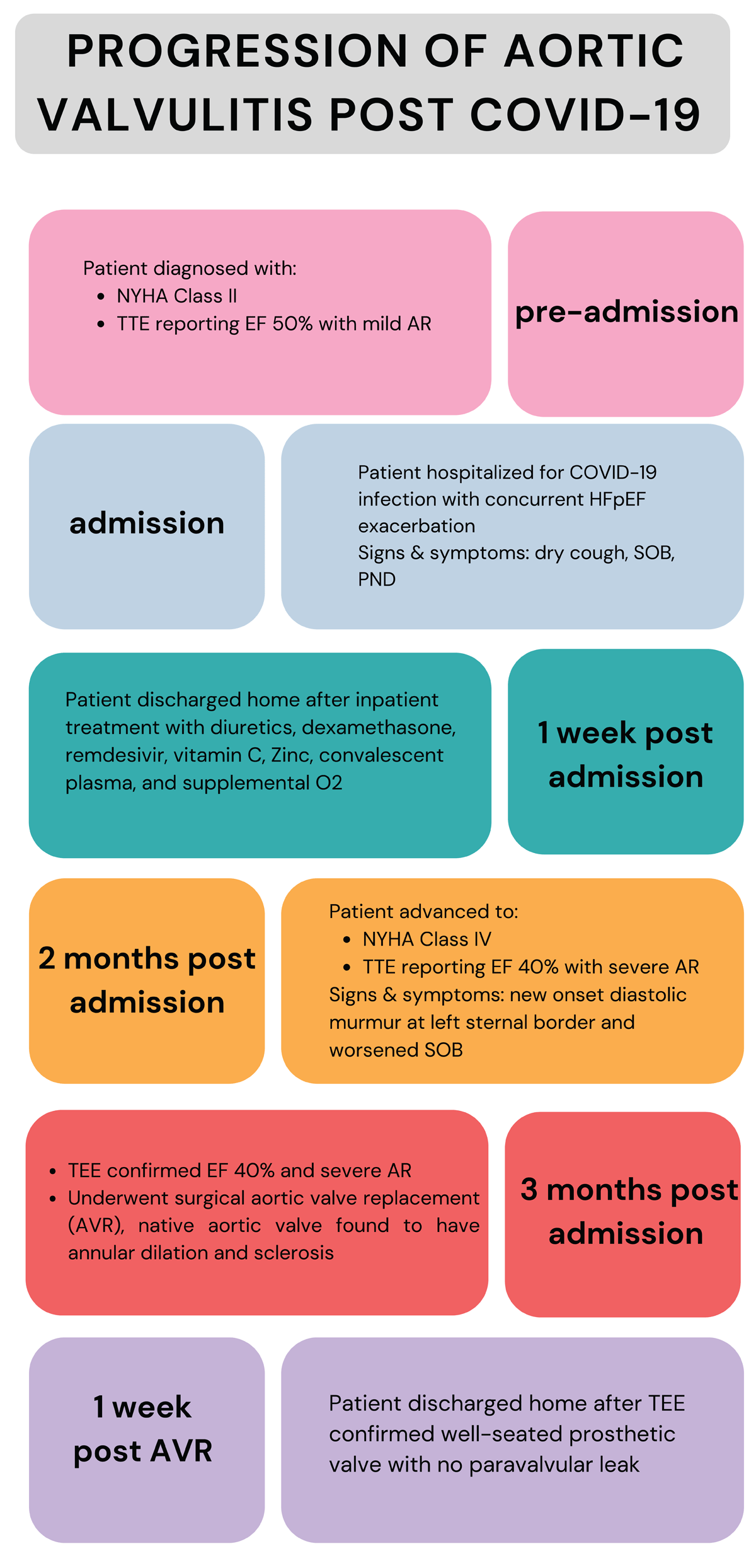
Figure 4
Progression of symptoms, workup, and management in patient with aortic valvulitis in the setting of COVID-19.
Table 1
Timeline of transthoracic echocardiogram findings in patient prior, during, and post COVID-19 infection. TTE: transesophageal echocardiogram; AV: aortic valve; LVEF: left ventricle ejection fraction; LV: left ventricle; AR: aortic regurgitation
| DATE OF STUDY | TTE 4/14/2019 | TTE 7/21/2020 | TTE 11/11/2020 *TIME OF COVID-19 INFECTION* | TTE 1/12/2021 | TEE 1/27/2021 *INTRA-OPERATIVE, DURING AV REPLACEMENT* | TTE 1/19/2022 | TTE 1/26/2023 | TTE 1/24/2024 | TTE 3/3/2025 |
|---|---|---|---|---|---|---|---|---|---|
| LVEF | 55 - 60% | 55 - 60% | 45 - 50% | 45% | 40 - 45% | 55% | 53.1% | 50% | 50 - 55% |
| LV diastolic diameter | 5.97 cm | 5.66 cm | 7.0 cm | 6.79 cm | 5.11 cm | 5.28 cm | 5.64 cm | 4.99 cm | |
| LV systolic diameter | 4.4 cm | 3.83 cm | 3.4 cm | 5.02 cm | 3.43 cm | 3.83 cm | 3.82 cm | 3.65 cm | |
| Left ventricle | Grade II diastolic dysfunction, normal LV systolic function | Normal diastolic and systolic LV function | Low-normal systolic LV function | Mild-moderate LV dilation, mild-moderate decrease in LV function | Diffuse hypokinesis, reduced LV systolic function | Grade I diastolic dysfunction, normal LV systolic function | Normal LV systolic and diastolic function | Grade I diastolic dysfunction, normal LV systolic function | |
| Aortic valve | Mild AR | Moderate AR | Moderate AR | Moderate-severe AR | Severe AR | Bioprosthetic AV, trace AR | Bioprosthetic valve, mild AR | Bioprosthetic valve, no valvular or peri-valvular AR | Bioprosthetic valve, mild AR |
Several etiologies for the high rates of cardiovascular complications associated with COVID-19 have been proposed, although the exact pathophysiologic mechanism has yet to be determined. Many of these theories are centered on the ACE2 receptor, which has been identified as the conduit for virus entry into the human body. This receptor is even more highly expressed in the heart than in the lungs, which is the target organ of this virus, suggesting susceptibility of the cardiovascular system to complications of COVID-19. Additionally, myocardial ACE2 expression was found to be significantly elevated in individuals with underlying heart failure such as this patient.7
Once the virus gains entry through ACE2, the receptor is subsequently downregulated leading to decreased conversion of angiotensin II (Ang-II) to angiotensin 1-7 (Ang 1-7).13 Ang-II exerts many detrimental effects on the cardiovascular system, including the promotion of fibrosis, proliferation, and inflammation. Conversely, Ang 1-7 is cardioprotective and counteracts these effects. The ACE2 receptor has been explicitly identified on the aortic valve cusps, rendering this valve susceptible to complications of COVID-19.8 Thus, it is plausible that this preference of the renin-angiotensin system (RAS) towards Ang-II stimulated by virus entry at the aortic valve can lead to degeneration of the valve and perivalvular apparatus. When the integrity of the valve is compromised, this can certainly manifest as valvular insufficiency as seen in this patient.
The ACE2 receptor was also found to be uniquely expressed in pericytes, a type of perivascular cell that is integral to maintaining endothelial cell function in the capillary vessels of the myocardium.7 Therefore, virus-infected pericytes can lead to capillary endothelial cell dysfunction and, ultimately, impairment in the microcirculation of the cardiovascular tissue. Not only can this manifest as troponin elevation and diffuse myocardial injury, it could also induce acute valvular insufficiency if the perivalvular pericytes and microcirculation are affected.14
Collectively, the proinflammatory state and microcirculatory dysfunction mediated by virus-infected ACE2 receptors and pericyte cells, respectively, in the setting of a systemic viral illness provides a credible etiology for ensuing valvular insufficiency. Thus, it is imperative to consider valvular disease, specifically that of the aortic valve, as one of the many cardiovascular complications of COVID-19. Furthermore, individuals with underlying heart failure are even more susceptible to a complicated disease course, in this case resulting in aortic insufficiency necessitating valve replacement.
Conclusion
Due to the systemic and debilitating nature of the COVID-19 virus, it is crucial that we understand its variable effects on the human body as well as the associated long-term complications. The cardiovascular system has been identified as one of the organ systems most vulnerable to complication due to the increased myocardial expression of the ACE2 receptor, which functions as the access point for virus entry into the human host. In particular, this receptor has also been identified on the aortic valve. We know that the RAS axis plays an integral role in the maintenance of valvular integrity. When this system is inclined towards inflammation by the virus, valvular pathology and hemodynamic alterations may rapidly ensue. In this patient, subacute AR and decompensated heart failure developed in less than 2 months following infection with COVID-19. This ultimately required open thoracic surgery and valvular replacement. Moving forward, it is necessary that we consider valvular disease, particularly that of the aortic valve, as a potential cardiovascular complication of COVID-19.
Key Points
In the setting of patients with a history of COVID-19 presenting with subacute aortic regurgitation and relatively rapid development of decompensated heart failure, complications of COVID-19 should be considered in the differential.
COVID-19, via manipulation of the angiotensin-converting enzyme 2 receptors and renin-angiotensin-aldosterone system pathway, can lead to aortic valvular inflammation and subsequent signs and symptoms consistent with aortic regurgitation.
Competing Interests
The authors have no competing interests to declare.