
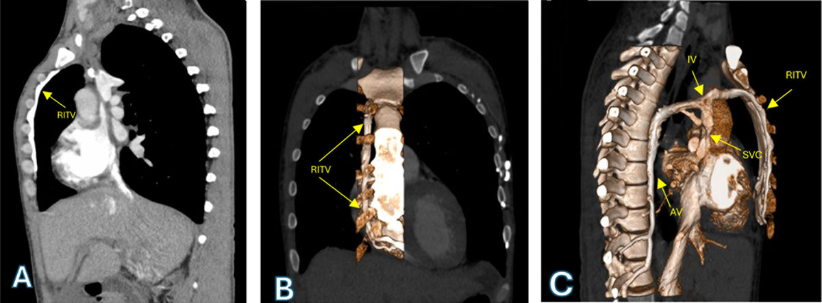
Figure 1
(A) Chest computed tomography angiography showing a dilated RITV. (B) 3D reconstruction showing the RITV in its parasternal course. (C) 3D reconstruction showing the remnant of the IV, the confluence of the AV, and the RITV in the SVC. RITV: right internal thoracic vein, IV: innominate vein, AV: azygos vein, SVC: superior vena cava
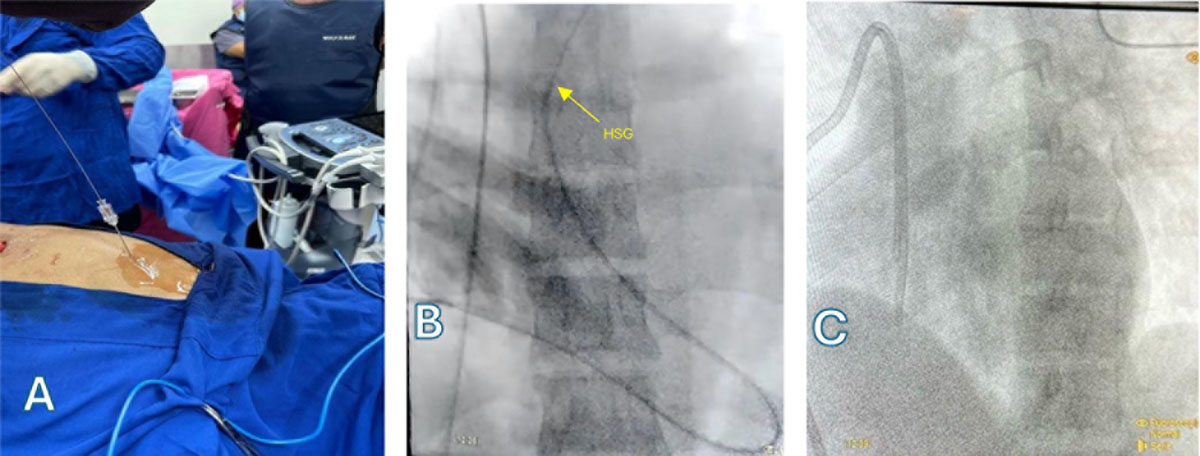
Figure 2
(A) Ultrasound-guided puncture of the right internal thoracic vein. (B) Passage of catheter and HSG to the pulmonary arteries. (C) Final position of the PDC. HSG: high-support guidewire, PDC: permanent dialysis catheter
Video 1
This video demonstrates the procedure from the passage of the introducer to the advancement and final positioning of the permanent dialysis catheter through the right internal thoracic vein; see also at https://youtu.be/3o-lr9zxoP8.