
Introduction
While patients with advanced chronic kidney disease depend on adequate vascular access for hemodialysis,1 complications such as dysfunction of prior arteriovenous fistulas and depletion of traditional catheter sites may necessitate the use of unconventional approaches.2 Computed tomography angiography (CTA), 3-dimensional reconstructions, and ultrasound can help identify alternative vascular pathways for catheter placement, ensuring continued hemodialysis treatment.3 In this case, we successfully achieved the percutaneous placement and tunneling of a 14F x 40 cm permanent dialysis catheter through the right internal thoracic vein, obtaining flows above 300 mL/min in the hemodialysis machine.
Case Presentation
We report a case of a 30-year-old patient with dysfunctional arteriovenous fistulas and a history of chronic renal failure KDOQI V diagnosed five years ago. Renal replacement therapy with catheters had been attempted throughout his history, including three right jugular catheters (two temporary and one tunneled); one temporary left jugular catheter; two peritoneal catheters presenting peritonitis in both cases; three right femoral catheters (one temporary and two tunneled); one temporary left femoral catheter; two right transcaval catheters; and one attempt at transcaval catheter placement via the left iliac vein. Finally, it was possible to partially place a left subclavian catheter that generated less than 180 mL/min, which provided suboptimal treatment that stopped working one day before the procedure. Using computed tomography angiography (CTA), 3-dimensional reconstruction, and ultrasound studies (Figure 1), the treatment team decided (prior to a more invasive treatment) to place a permanent hemodialysis catheter 14F × 40 cm through the internal thoracic vein (ITV) using a percutaneous approach to successfully advance the catheter (Figure 2, Video 1).
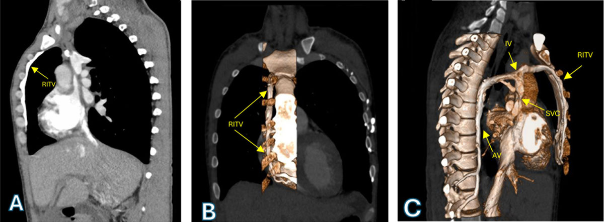
Figure 1
(A) Chest computed tomography angiography showing a dilated RITV. (B) 3D reconstruction showing the RITV in its parasternal course. (C) 3D reconstruction showing the remnant of the IV, the confluence of the AV, and the RITV in the SVC. RITV: right internal thoracic vein, IV: innominate vein, AV: azygos vein, SVC: superior vena cava
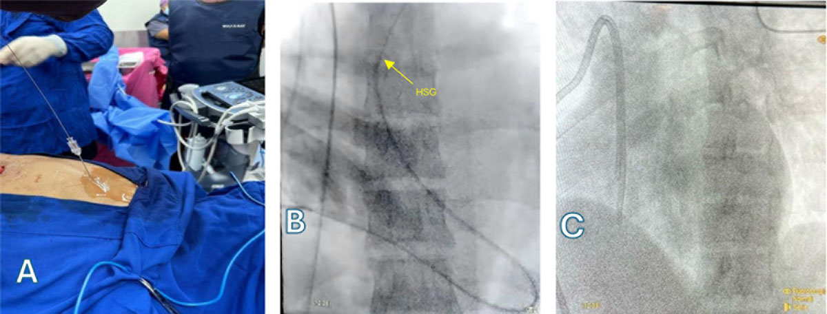
Figure 2
(A) Ultrasound-guided puncture of the right internal thoracic vein. (B) Passage of catheter and HSG to the pulmonary arteries. (C) Final position of the PDC. HSG: high-support guidewire, PDC: permanent dialysis catheter
Video 1
This video demonstrates the procedure from the passage of the introducer to the advancement and final positioning of the permanent dialysis catheter through the right internal thoracic vein; see also at https://youtu.be/3o-lr9zxoP8.
Discussion
Vascular access is paramount for the survival of hemodialysis patients. However, we often encounter situations where these accesses become exhausted due to thrombosis, infection, or simply wear and tear over time. In such cases, the urgent need to initiate or continue hemodialysis presents a significant challenge to the healthcare team.
The ITV, also known as the internal mammary vein, has emerged as a viable option for hemodialysis catheter placement, particularly when other vascular access sites have been exhausted or are inadequate.4 Its deep location, adequate caliber, and relative ease of access make it an attractive alternative, although not without risks and challenges. It also has been utilized for central venous catheter placement in both adults and children.5
The ITV descends along the medial border of the first costal cartilage and is protected by the ribs. Its size and blood flow vary among individuals, but in general, it offers a suitable caliber for hemodialysis catheter placement. The distance from the sternum to ITV gradually decreases as the vessel passes caudally. The diameter of the vessel similarly decreases along the vein’s caudal course.6
Placement of an ITV catheter is typically performed under ultrasound guidance. An incision is made in the skin, and a subcutaneous tunnel is dissected for the catheter. The distal end of the catheter is introduced into the ITV and secured to the skin.
Conclusion
Occasionally, patients present with exhausted vascular access, putting their lives at risk. This necessitates the use of unconventional or poorly described procedures to obtain a suitable pathway for hemodialysis catheter advancement.7 These procedures remain one of the most significant challenges faced by dialysis units, thus a multidisciplinary approach is essential.8
Competing Interests
The authors have no competing interests to declare.