
Question 1
Consider the options and find the answer in this video quiz, also at https://youtu.be/ovI9QbgoeWk.
Question 2
Consider the options and find the answer in this video quiz, also at https://youtu.be/ZTOQ5kgHjA8.
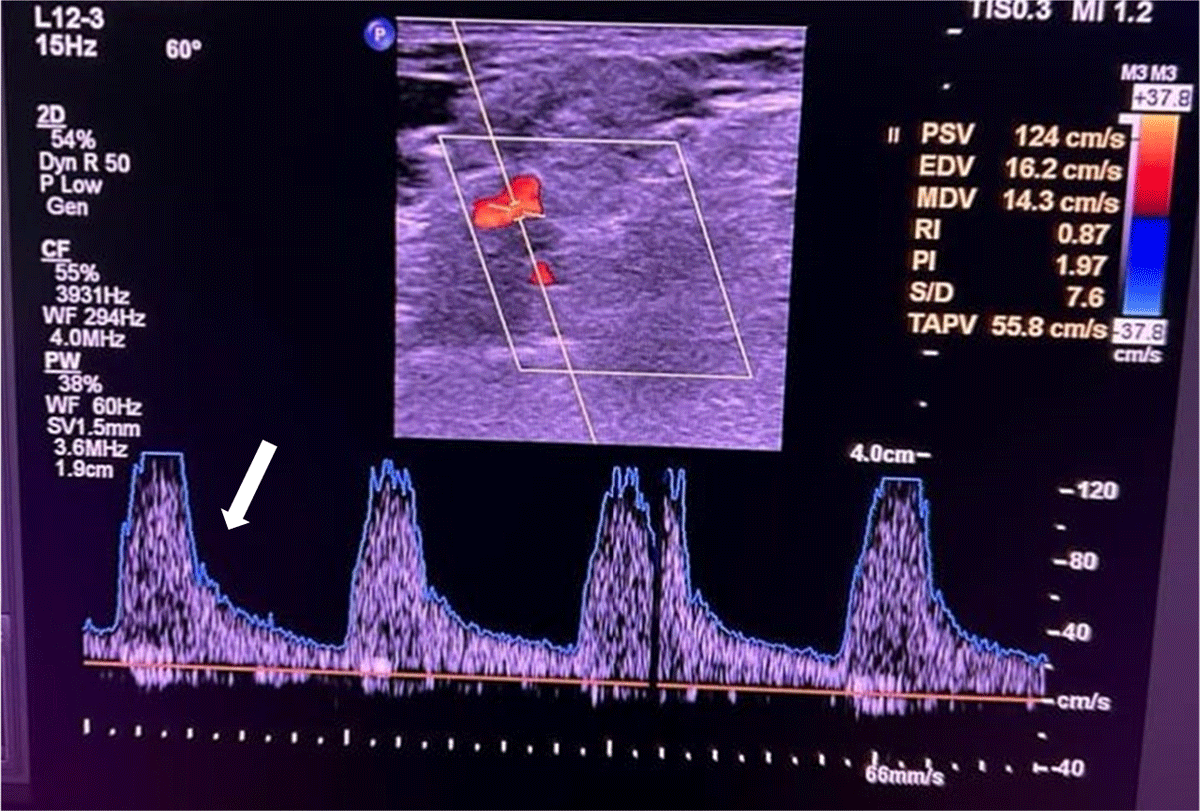
Figure 1
Ultrasound Doppler of left subclavian artery shows high peak velocity (white arrow).
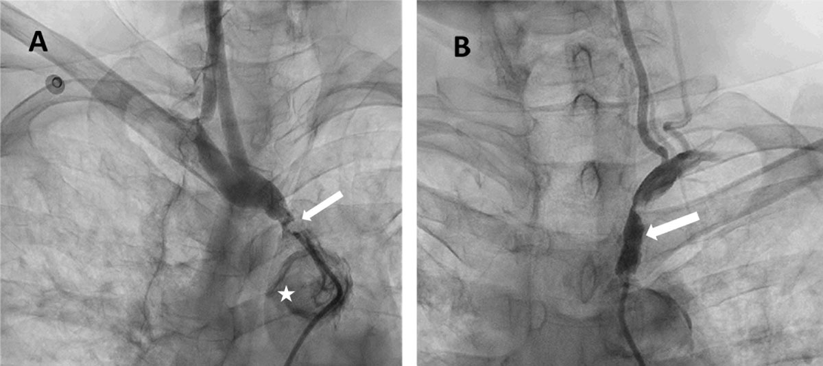
Figure 2
Bilateral subclavian artery stenosis shows (A) right innominate artery stenosis (white arrow) with calcific arch of aorta (star) and (B) left subclavian artery stenosis (white arrow).
Video 1
Angiogram of right innominate artery; see also at https://youtube.com/shorts/SFg8qFyH6KE.
Video 2
Angiogram of left subclavian artery; see also at https://youtube.com/shorts/0N23kWKQFjo.
Video 3
Angiogram of post stent flow; see also at https://youtube.com/shorts/YU8q_nEOPYo.
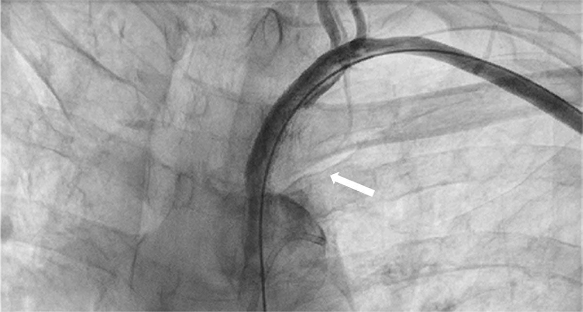
Figure 3
Percutaneous transluminal angioplasty of left subclavian artery with a 7-mm × 39-mm peripheral stent (white arrow).
Question 3
Consider the options and find the answer in this video quiz, also at https://youtu.be/3UBaja7_psw.