
Case Presentation
A male patient in his late 60s presented to the emergency triage of a tertiary care hospital with complaints of transient blurring of vision lasting for 20 minutes and was later identified to be in shock. His systolic blood pressure (SBP) was 60 mm Hg in both arms, and he was immediately started on fluids and inotropic support. He had a history of type 2 diabetes mellitus on insulin injection, untreated hypertension, and an unclear cardiac condition managed with herbal medicine. He also reported an old cerebrovascular accident/transient ischemic attack (TIA) on medical management for 6 years and percutaneous peripheral angioplasty of the right superficial femoral artery in 2016.
Question 1
What is the most likely cause of his presentation, and what would be the next best step for management?
Cardiogenic shock – initiate inotropic support
Hypovolemic shock – administer fluids
Acute aortic dissection – urgent imaging and surgical consult
Acute cerebrovascular accident (CVA) – urgent magnetic resonance imaging (MRI) of brain and neurology consultation
None of the above
Question 1
Consider the options and find the answer in this video quiz, also at https://youtu.be/ovI9QbgoeWk.
The patient was admitted to the cardiology intensive care unit (CICU) for further management of shock and to identify the cause. He was simultaneously assessed for consciousness level, lactate levels, renal parameters, and urine output. Troponin T levels, electrocardiography (ECG), and echocardiography were conducted to rule out a cardiac cause for shock. A neurology consultation also was sought to rule out acute CVA and/or TIA. Meanwhile, the patient continued to remain hypotensive despite the fluid challenge and inotropic support. The following findings were reported:
Consciousness levels: alert and oriented
Vitals:
Heart rate – 65 bpm
Bilateral arm systolic blood pressure – 60 mm Hg
Respiratory rate – 18/min
ECG: sinus rhythm, left ventricular (LV) hypertrophy
Echocardiography: moderate LV dysfunction with an ejection fraction of 38% and global hypokinesia of LV
MRI of the brain: acute focal infarct in the right peri trigonal white matter. Magnetic resonance angiogram revealed a complete occlusion of the left internal carotid artery and partial occlusion of the right internal carotid artery, with no focal neurological deficits.
His laboratory findings revealed the following:
Lactate: 4 mmol/L
Urea: 21 mg/dL
Creatinine: 1.26 mg/dL
Serial troponin T (in a gap of 6 hours):
Troponin T (1st sample): 0.016 ng/L
Troponin T (2nd sample): 0.020 ng/L
Blood counts:
Leukocytes: 8,900/mm3
Platelets: 2,50,000/mm3
Question 2 What clue leads to a correct diagnosis of this type of shock?
Feeble pulses
Moderate left ventricular dysfunction on echocardiography
Infarct on MRI
Normal lactate levels
Question 2
Consider the options and find the answer in this video quiz, also at https://youtu.be/ZTOQ5kgHjA8.
The patient’s normal lab values, refractory response to inotropes, and past medical history prompted a focused clinical examination, which revealed bilateral subclavian bruit. An emergency Doppler was performed (Figure 1) and showed a high peak velocity in both subclavian arteries, indicating high turbulence. Ultrasound Doppler of the vertebral artery did not show any signs of flow reversal. The patient was immediately weaned off inotropic support over the next 24 hours. Furthermore, because of the multiple risk factors with LV dysfunction, a cerebral four-vessel angiography and coronary angiography were planned. While securing the femoral arterial line, the invasive blood pressure showed a reading of 220/110 mm Hg.
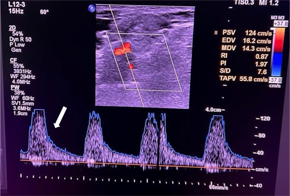
Figure 1
Ultrasound Doppler of left subclavian artery shows high peak velocity (white arrow).
Coronary angiography showed calcific multivessel disease, and four-vessel angiography confirmed bilateral subclavian artery stenosis (SAS) (Figure 2; Videos 1, 2). The patient underwent percutaneous transluminal angioplasty of the left subclavian artery with a 7-mm × 39-mm stent, tremendously aiding in the assessment and management of hypertension (Figure 3; Video 3).
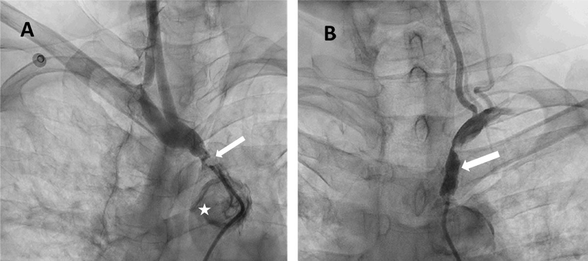
Figure 2
Bilateral subclavian artery stenosis shows (A) right innominate artery stenosis (white arrow) with calcific arch of aorta (star) and (B) left subclavian artery stenosis (white arrow).
Video 1
Angiogram of right innominate artery; see also at https://youtube.com/shorts/SFg8qFyH6KE.
Video 2
Angiogram of left subclavian artery; see also at https://youtube.com/shorts/0N23kWKQFjo.
Video 3
Angiogram of post stent flow; see also at https://youtube.com/shorts/YU8q_nEOPYo.
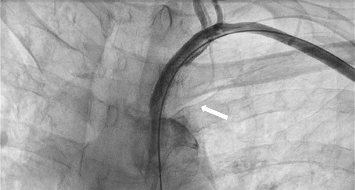
Figure 3
Percutaneous transluminal angioplasty of left subclavian artery with a 7-mm × 39-mm peripheral stent (white arrow).
Post angioplasty, the patient was started on dual antiplatelet therapy and statin medications and was given intravenous nitroglycerine infusion followed by oral antihypertensive agents to control accelerated hypertension. However, this induced a sudden left-sided weakness due to probable ischemia in watershed territory. Thus, treatment was next directed towards achieving a balance between managing hypertension and preventing cerebral hypoperfusion as well as treatment of remaining comorbidities—therefore, the dosage of oral antihypertensives was titrated to maintain a target SBP of 140 mm Hg. This stabilized his neural deficits, and he was referred to physiotherapy to facilitate functional recovery. Future interventions will look at revascularization of the right subclavian artery, right innominate artery, and coronaries after recovery from acute stroke.
Question 3 What is the most likely diagnosis?
Left subclavian artery stenosis
Bilateral subclavian artery stenosis
Vertebral steal syndrome
All of the above
Question 3
Consider the options and find the answer in this video quiz, also at https://youtu.be/3UBaja7_psw.
Discussion
This case report highlights a unique patient with extensive peripheral vascular disease with bilateral SAS presenting as pseudo-shock. SAS is reported in approximately 1.9% of the community population with peripheral artery disease, only half of whom are expected to have coronary artery disease and one-third to have vertebral artery and carotid artery involvement.1,2 Furthermore, pseudo-shock is one of the rarest presentations of severe bilateral SAS, which makes it challenging to diagnose.2, 3, 4, 5, 6 Guidelines recommend using bilateral brachial artery blood pressure measurements to diagnose upper extremity artery involvement, as well as noting a difference of 15 mm Hg in the SBP measurement between the two upper extremities, to raise the suspicion of bilateral SAS.3 This was not seen in our case due to the bilateral subclavian artery involvement. This patient had polyvascular disease and presented with “refractory shock” along with bilateral subclavian bruit on examination that led one to suspect bilateral SAS, similar to some other case reports.2,4, 5, 6
A coronary angiogram and cerebral four-vessel angiogram were done due to the patient’s multiple risk factors and to explore the cause of shock in the presence of LV dysfunction, which aided in the diagnosis of pseudo-shock. Revascularization of the left subclavian artery further facilitated hypertension management by resulting in an accurate blood pressure, making it a primary choice of intervention.7
The patient has a long route to recovery and was discharged after obtaining optimal control over his symptoms and his disease condition along with managing his multiple comorbidities. Due to the upper extremity arterial involvement, following coronary artery bypass grafting, the patient carries a risk of vertebrobasilar steal syndrome. Keeping this in mind, the way forward is percutaneous intervention of the right subclavian artery, right innominate artery, and coronaries after recovery from acute stroke, in line with the 2017 European Society of Cardiology guidelines.8
Conclusion
Bilateral SAS is rare in prevalence and known for its diagnostic challenge. This case report shows the possible diagnosis of pseudo-shock for a patient having polyvascular disease with bilateral SAS. Although he initially presented with neurological symptoms and was suspected to be in shock, it finally was determined to be a case of bilateral SAS with “pseudo-shock.” During the revascularization of the left subclavian artery, the femoral intra-arterial blood pressure revealed him to have accelerated hypertension instead. This case highlights the importance of thoroughly evaluating various vascular territories in a patient with polyvascular disease.
Key Points
Shock that is refractory to its symptoms can help to rule out pseudo-shock by screening lactate levels and renal parameters.
Thorough evaluation of other vascular territories is required in patients with atherosclerosis to predict and prevent polyvascular events.
This case emphasizes the critical need to maintain blood pressure within the optimal range to avert ischemic events in watershed areas, even post-revascularization.
CME Credit Opportunity
Houston Methodist is accredited by the Accreditation Council for Continuing Medical Education (ACCME) to provide continuing medical education for physicians.
Houston Methodist designates this enduring material for a maximum of 1 AMA PRA Category 1 Credit™. Physicians should claim only the credit commensurate with the extent of their participation in the activity. Click to earn CME credit: https://learn.houstonmethodist.org/MDCVJ-21.1.
Competing Interests
The authors have no competing interests to declare.