
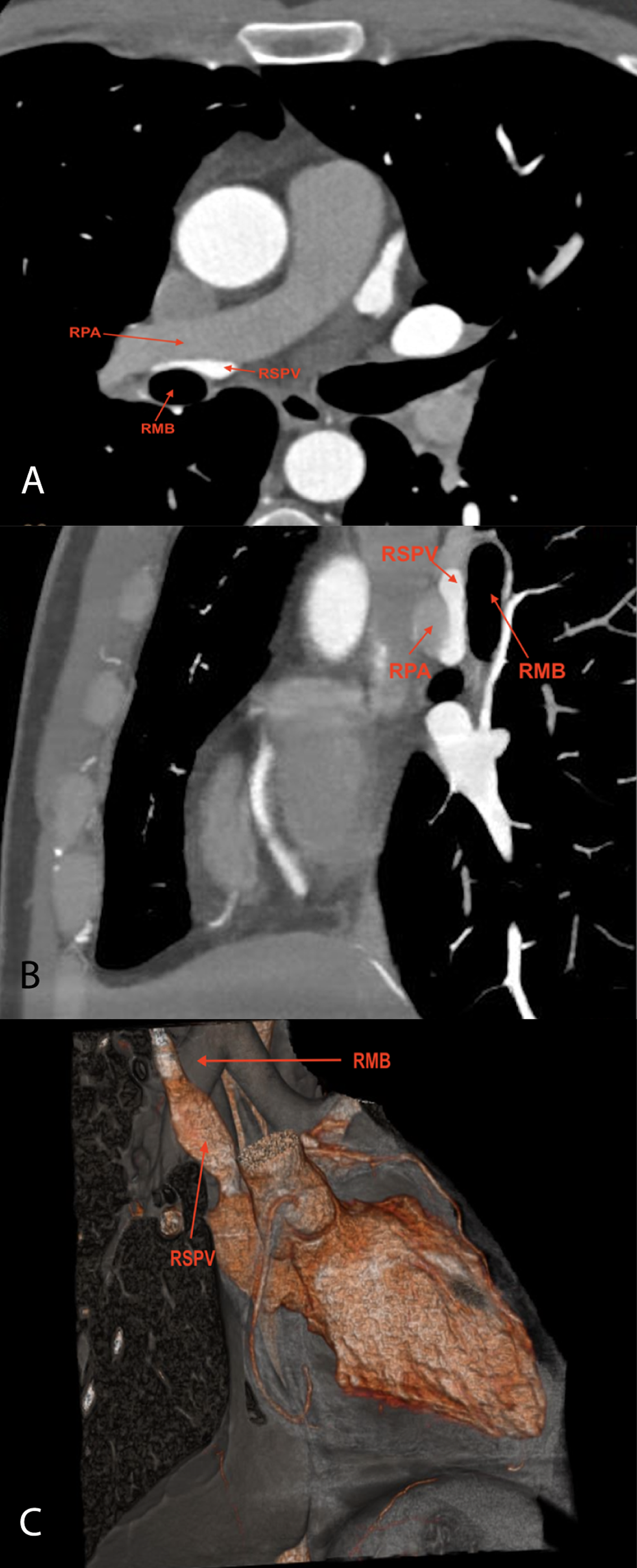
Figure 1
(A) A contrast-enhanced axial chest computed tomography (CT) scan shows the right superior pulmonary vein (RSPV) is aberrantly coursing between the right pulmonary artery (RPA) and the right main bronchus (RMB), and it is compressed in this anatomical configuration. (B) A contrast-enhanced chest CT image shows the RSPV compressed between the RPA and RMB. (C) A 3-dimensional CT image shows the RSPV anterior to the RMB and flattened due to compression by the RPA and RMB.
Video 1
A contrast-enhanced axial chest computed tomography scan shows that the right superior pulmonary vein is aberrantly coursing between the right pulmonary artery and the right main bronchus; see also at https://youtube.com/shorts/qC4w2flCJlM.