
Case Presentation
A previously healthy 35-year-old male patient with a smoking history (30 packs per year) presented with atypical left-sided chest pain and left forearm numbness. He did not have symptoms of effort-related chest pain, dyspnea, palpitations, hemoptysis, or syncope. The patient works as a contractor and does not engage in regular physical activities. Upon physical examination, his vital signs, heart sounds, and bilateral breath sounds were unremarkable.
The initial diagnostic workup included an electrocardiogram, which revealed no significant abnormalities, and a chest x-ray that was unremarkable. Two-dimensional transthoracic echocardiography demonstrated normal structure and function of all four cardiac chambers and valves, without evidence of intracardiac shunts, masses, pulmonary arterial hypertension, or pericardial effusion.
Coronary computed tomography angiography showed unremarkable coronary arteries. However, it revealed compression and flattening of the right superior pulmonary vein due to its aberrant course between the right pulmonary artery and right main bronchus (Figure 1; Video 1).
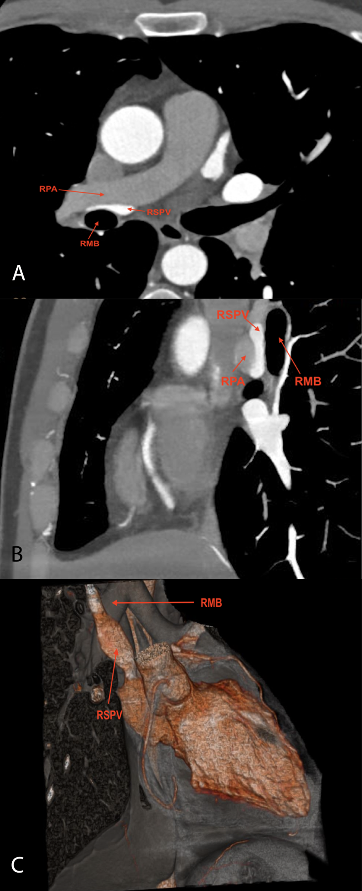
Figure 1
(A) A contrast-enhanced axial chest computed tomography (CT) scan shows the right superior pulmonary vein (RSPV) is aberrantly coursing between the right pulmonary artery (RPA) and the right main bronchus (RMB), and it is compressed in this anatomical configuration. (B) A contrast-enhanced chest CT image shows the RSPV compressed between the RPA and RMB. (C) A 3-dimensional CT image shows the RSPV anterior to the RMB and flattened due to compression by the RPA and RMB.
Video 1
A contrast-enhanced axial chest computed tomography scan shows that the right superior pulmonary vein is aberrantly coursing between the right pulmonary artery and the right main bronchus; see also at https://youtube.com/shorts/qC4w2flCJlM.
Our case revealed no changes in the lung tissue, including septal thickening or ground-glass opacities. Additionally, the right ventricle was normal. Consequently, the management strategy was conservative, involving serial echocardiography follow-ups and close monitoring.
Competing interests
The authors have no competing interests to declare.