
Introduction
Three-dimensional (3D) printing technology has been increasingly used in medical procedures to aid anatomical understanding and preprocedural planning. We report the use of 3D printing for catheter ablation of an atrial tachycardia in a patient with congenital heart disease with an unusual cardiac anatomy.
Case
A 45-year-old male with recurrent symptomatic supraventricular tachycardia (SVT) was referred to our center for an electrophysiology study (EPS) and ablation. Past medical history included secundum atrial septal defect (ASD) with a prior patch closure, SVT, atrial flutter (AFL), and atrial tachycardia (AT). The patient had prior ablation procedures for SVT, AT, and AFL. Baseline echocardiography showed left ventricular ejection fraction of 50%, enlarged left atrium, no residual ASD, and otherwise unremarkable findings.
With intravenous isoproterenol infusion, SVT with variable tachycardia cycle length (TCL) of 310 ms to 340 ms was induced by atrial pacing (Figure 1A). The tachycardia had a 1:1 atrioventricular (AV) relationship and variable ventriculoatrial intervals. The septal atrial activation was high to low. Changes in atrial activation interval preceded changes in ventricular activation interval. Ventricular overdrive pacing resulted in AV dissociation and did not affect the atrial TCL (Figure 1B). Atrial overdrive pacing at different pace cycle lengths from the same location resulted in variable post-pacing intervals and no evidence of progressive fusion. The tachycardia spontaneously terminated with a ventricular signal. These findings were consistent with a focal AT.
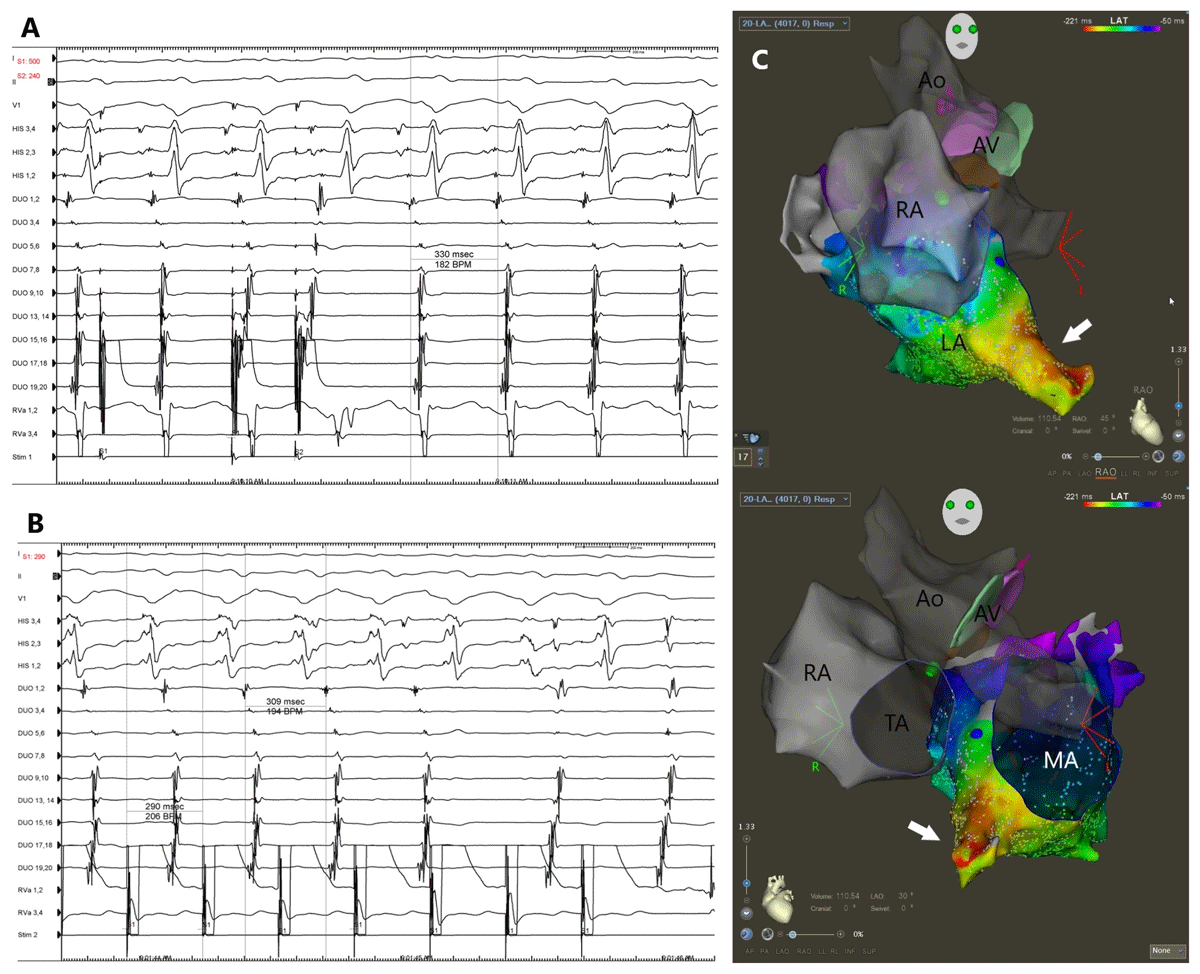
Figure 1
(A) Supraventricular tachycardia (SVT) with variable tachycardia cycle length (TCL) from 310–340 ms was induced by atrial pacing. There was a 1:1 atrioventricular relationship with long RP intervals. Septal atrial activation was high to low. (B) Ventricular overdrive pacing resulted in atrioventricular dissociation without affecting the tachycardia (SVT spontaneously terminated during pacing). (C) Three-dimensional electroanatomic activation mapping during atrial tachycardia in the right anterior (top) and left anterior (bottom) oblique views. The earliest site of atrial activation was located in a diverticulum located on the anterior-inferior aspect of the left atrium (white arrows). Panels A and B depict electrocardiographic leads I, II, and V1; electrograms of a quadripolar catheter in the His position (HIS 1,2 through 3,4); electrograms of a PENTARAY diagnostic catheter (labeled as DUO 1,2 through DUO 19,20); and electrograms of right ventricular apex (RVa 1,2 and 3,4). The PENTARAY diagnostic catheter was positioned in the high right atrium. Ao: ascending aorta; AV: aortic valve; LA: left atrium; MA: mitral annulus; RA: right atrium; TA: tricuspid annulus
3D electroanatomic activation mapping during AT was created using a PENTARAY® catheter with the CARTO™ mapping system (Biosense Webster, Johnson & Johnson) and showed the earliest site of atrial activation in a diverticulum-like structure located on the anterior-inferior aspect of the left atrium (Figure 1C). Due to an unusual anatomy, catheter ablation was not performed out of concern for safety. Atrial tachycardia spontaneously terminated after cessation of isoproterenol infusion. It was also noted that the coronary sinus could not be cannulated by a catheter during the procedure. The procedure was concluded.
Cardiac computed tomography (CT) with intravenous contrast was obtained to further define the anatomy. Cardiac CT showed a large left atrial diverticulum (4 cm in length and 2 cm in diameter at the ostium) located on the anterior-inferior aspect of the left atrium that tapered and terminated into the perihepatic fat tissue (Figure 2A). The coronary sinus drained directly into the left atrium. It was suggested that 3D models would be helpful for pre-ablation planning by providing realistic perspectives of the diverticulum in relation to the nearby structures and improving spatial orientation of the operators and support staff.
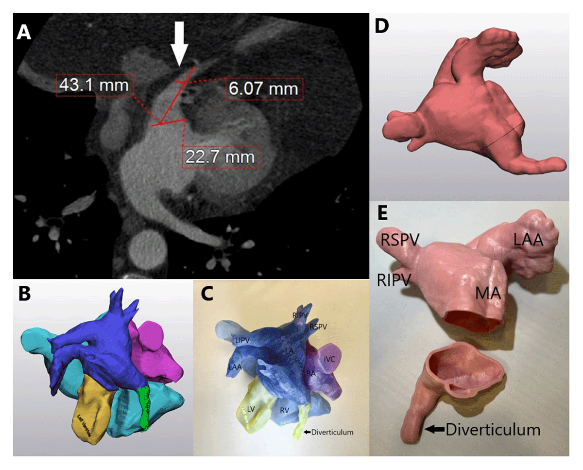
Figure 2
(A) Cardiac computed tomography showed a large left atrial diverticulum (4 cm by 2 cm at the ostium) located on the anterior-inferior aspect of the left atrium that tapered and terminated into the perihepatic fat tissue (white arrow). (B, C) Digital and 3-dimensional (3D) printed models show the left atrium diverticulum in relationship with other cardiac structures. (D, E) Digital and 3D printed models of the left atrium with the inferior portion transected to show left atrium diverticulum in relationship with other left atrial structures. IVC: inferior vena cava; LA: left atrium; LAA: left atrial appendage; LIPV: left inferior pulmonary vein; LV: left ventricle; MA: mitral annulus; RA: right atrium; RIPV: right inferior pulmonary vein; RSPV: right superior pulmonary vein; RV: right ventricle
3D printing technology was used to create models of the heart. The first model depicted each chamber of the heart (Figure 2B), and the second model depicted the left atrium that was transected to show the opening of the diverticulum with a cross-sectional view (Figure 2D). These digitally created models were then printed by a 3D printer into physical models (Figure 2C, 2E). Detailed processes of the creation of 3D printed models can be found below in the section on “Creation of 3D Printed Models.”
With better understanding of the anatomy from the cardiac computed tomography (CT) and 3D models, catheter ablation in the diverticulum was deemed technically feasible with the aid of a steerable sheath without excessive risk of collateral damage to the nearby structures. A repeat EPS was performed. An AT with TCL of 370 ms was induced. A transseptal puncture was obtained, and a steerable sheath was placed across the interatrial septum. Activation mapping showed the earliest site near the ostium of the left atrial diverticulum (Figure 3A). Radiofrequency ablation at the earliest site of activation resulted in termination of the AT and initiation of a different AT with a TCL of 315 ms. The second AT was mapped earliest inside the diverticulum (Figure 3B). Ablation in this area resulted in termination of the second AT. There was no further inducible arrhythmia. The patient tolerated the procedure well without any complications. A 6-month follow-up visit showed no clinical recurrence of AT.
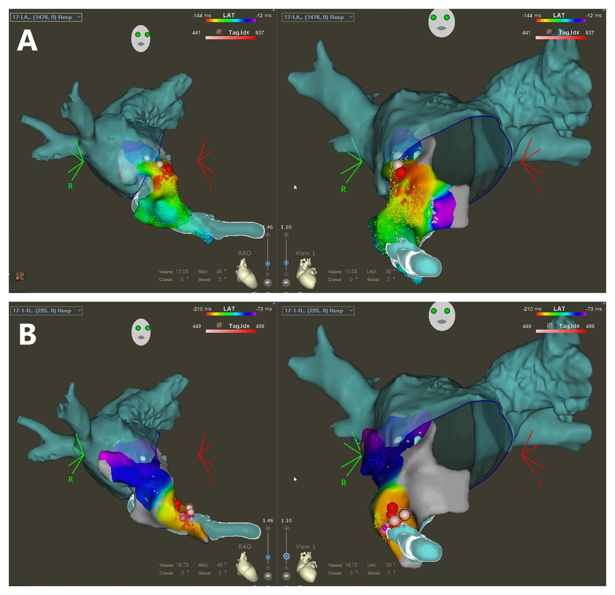
Figure 3
Three-dimensional electroanatomic activation mapping overlaying CT reconstructed images of the left atrium during atrial tachycardia (AT). (A) During the first AT with tachycardia cycle length (TCL) 370 ms, AT was mapped to the earliest site near the ostium of the left atrial diverticulum. Ablation resulted in termination of the first AT and initiation of the second AT. (B) During the second AT with TCL 315 ms, AT was mapped to the earliest site inside the diverticulum. Ablation at the site resulted in termination of the second AT. Left panels: right anterior oblique view; right panels: left anterior oblique view
Discussion
Extending structures from the left atrium have been described in different terms such as accessory appendage, diverticulum, pouch, or aneurysm.1 A study of cardiac CT showed the prevalence of left atrial diverticula, left atrial pouch, and left atrial accessory appendage of 28.4%, 24%, and 10%, respectively.2 There was no difference in the prevalence of these structures in patients with and without atrial fibrillation.2 The abnormal structure in this case was located on the anterior-inferior aspect of the left atrium, which then tapered and terminated in the perihepatic area. This raises the possibility that this structure could be partial anomalous hepatic venous drainage to the left atrium. Anomalous hepatic venous drainage to the left atrium is an uncommon entity, with sporadic case reports in the literature.3 It has been reported in patients with atrial septal defect.4
Arrhythmias are common in adult congenital heart disease (ACHD), affecting 15% of patients with this condition.5 Atrial fibrillation, isthmus-dependent typical atrial flutter, and intra-atrial reentrant tachycardia (IART) are the most common types of atrial arrhythmia in ACHD.6 Atrial arrhythmia involving atrial diverticula is exceedingly rare, with only a few reports of catheter ablation of right atrial diverticulum-related accessory pathways7,8 and left atrial diverticulum related macrorentrant AT.9 To the best of our knowledge, there has been no report of focal atrial tachycardia originating from a left atrial diverticulum as in this case. Focal atrial tachycardia is less common than IART in ACHD but has a higher success rate with catheter ablation.10
3D printing has been increasingly used in the medical field, mainly involving surgical procedures.11 In cardiology, 3D printing has been used to aid in anatomical understanding and preprocedural planning in congenital heart disease, left atrial appendage closure, left ventricular pseudoaneurysm, and valvular heart disease.12 The use of 3D printing in cryoballoon ablation of atrial fibrillation was studied and showed decreased contrast use compared to routine standard of care.13
3D printed models can be created using data from cardiac topography, magnetic resonance imaging, or echocardiogram. Although 3D printing can potentially improve procedural success rate and decrease complications, this has not been shown in large controlled studies.12 3D printed physical models provide more realistic perceptions of the anatomy compared to cross-sectional images or virtual reconstruction. The models potentially improve spatial orientation of the operators and support staff and may be used to provide education to medical trainees, healthcare providers, and patients. Additionally, 3D printing comes with added costs that can be analyzed and incorporated into the healthcare reimbursement system.14 The costs of these models vary by complexity; for example, the 3D lab at our institution generally charges a minimum of $3,500 per model. The personnel time and overhead costs for the staff, equipment, software, and space are much more significant than the material consumed.
Creation of 3D Printed Models
3D CAD File Preparation
Materialise Mimics Innovation Suite (Materialise) software was utilized for segmentation and digital preparation of the 3D printed model. Digital Imaging and Communications in Medicine (DICOM) files were imported and uploaded into the Mimics software. The computed tomography had a slice thickness of 0.625 mm and pixel spacing was 0.462891 mm. Custom thresholding was utilized, as there is no preset threshold in the software for cardiac tissue. Each chamber of the heart was segmented separately, using manual segmentation to include cardiac tissue that did not fall within the threshold and exclude artifact which had been incorrectly included. This then resulted in an initial raw 3D computer-aided design (CAD) model of the patient’s heart, with each chamber of the heart modeled as a separate part (Figure 4).
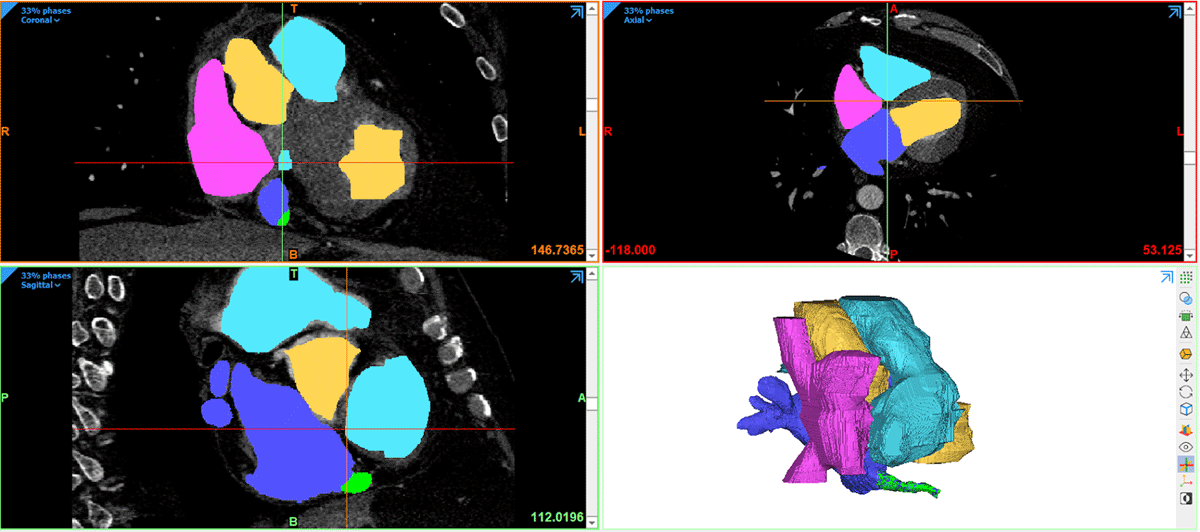
Figure 4
Initial raw 3-dimensional computer-aided design model of the patient’s heart, with each chamber of the heart modeled as a separate part.
Next, the unsculpted CAD model was exported as standard triangle language (STL) files to Materialise 3-Matic (Materialise) for digital sculpting and cleaning of each STL part to fill any holes present on the model, remove internal geometries which may cause failure during printing, and digital smoothing to remove any sharp edges.
3D Printing
Once the initial digital sculpting and cleaning was complete, two different models were created to best portray the relevant anatomy. In the first model (Figure 5), each chamber of the heart and the diverticulum would be colored differently, semitranslucent, and labeled. This model also included labels of the opening diameter and length of the diverticulum. To create this model, digital measurements of the diverticulum opening diameter and length were taken using Materialise 3-Matic. Then, using the Materialise 3-Matic “Quick Label” feature, each chamber of the heart, as well as the measurements taken of the diverticulum, were added to the 3D printed model as a text label. Once labels had been added, each “part” of the model was selected and exported as a 3D manufacturing format (3 mf) file to GrabCAD Print software (Stratasys). Once in GrabCAD, each part was assigned a different color, and translucency for each was set to 50%.
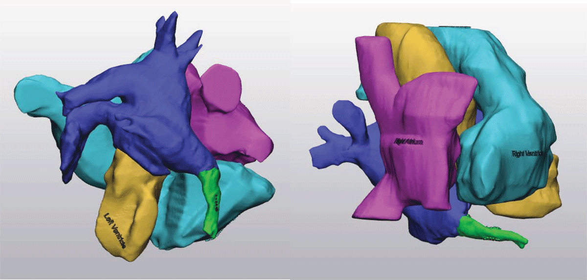
Figure 5
The first model portrays each chamber of the heart with the diverticulum colored differently, semitranslucent, and labeled. The measurements of the diverticulum are added to the 3-dimensional printed model as a text label.
The second model (Figure 6) would be hollow and opaque with a digitally created cut down the middle, so that the cardiac team could visualize the opening of the diverticulum from the inside of the heart. The digitally sculpted and cleaned parts were merged to create one file containing the whole model of the heart. Then, this model was wrapped and hollowed. Finally, using the 3-Matic interactive cut feature, a digital cut was created so the cardiovascular team could visualize the opening to the diverticulum. Lastly, using the 3-Matic “pins and holes” feature, pins and holes were added so that the model could be placed together as a whole heart or opened to visualize the internal anatomy. This was then exported as a 3 mf file to the GrabCAD software, where it was assigned a color.
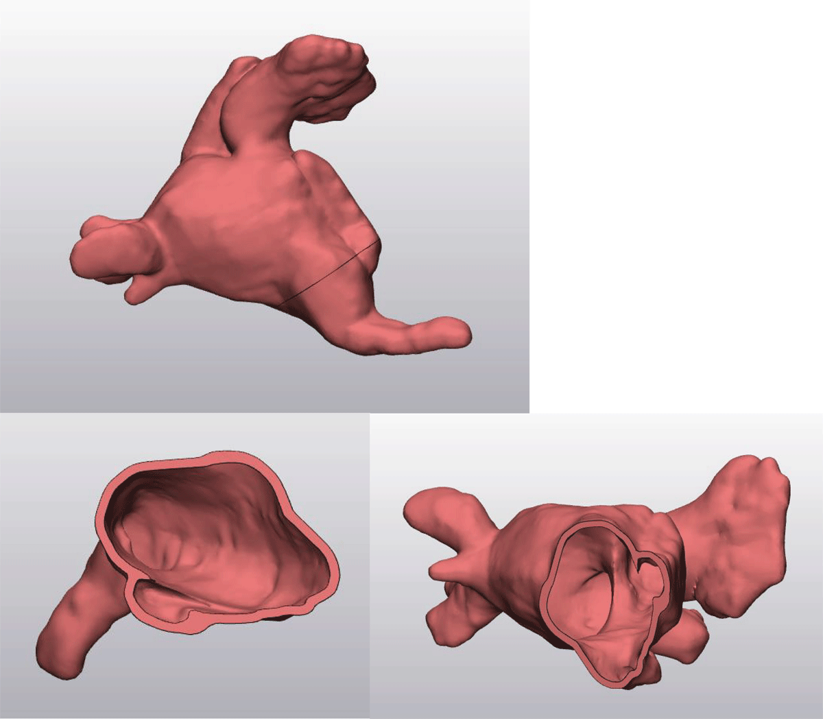
Figure 6
The second model is hollow and opaque with a digitally created cut down the middle, so that the cardiac team can visualize the opening of the diverticulum from the inside of the heart.
These models were then printed simultaneously on the Stratasys J5 Medijet printer (Stratasys). Models were post-processed including pressurized water for support material removal and an isopropanol rinse to clean residue. Models were cleaned following the J5 guidelines and delivered to the cardiac cath lab for use by the clinical team.
Conclusion
We report a rare case of focal atrial tachycardia originating from a large left atrial diverticulum of unclear embryonic origin in a patient with coronary sinus drainage to the left atrium and history of repaired secundum ASD. Due to the complex and unfamiliar anatomy, 3D printing was used for preprocedural planning for follow-up procedure resulting in successful ablation of the tachycardia without complications.
Competing Interests
The authors have no competing interests to declare.