
Have a personal or library account? Click to login
Retroesophageal Left Brachiocephalic Vein with Right-Sided Aortic Arch in Tetralogy of Fallot: A Rare Venous Anomaly Demonstrated on Computed Tomography Angiography Cover

Retroesophageal Left Brachiocephalic Vein with Right-Sided Aortic Arch in Tetralogy of Fallot: A Rare Venous Anomaly Demonstrated on Computed Tomography Angiography

Open Access
|Nov 2024

Figures & Tables

Figure 1

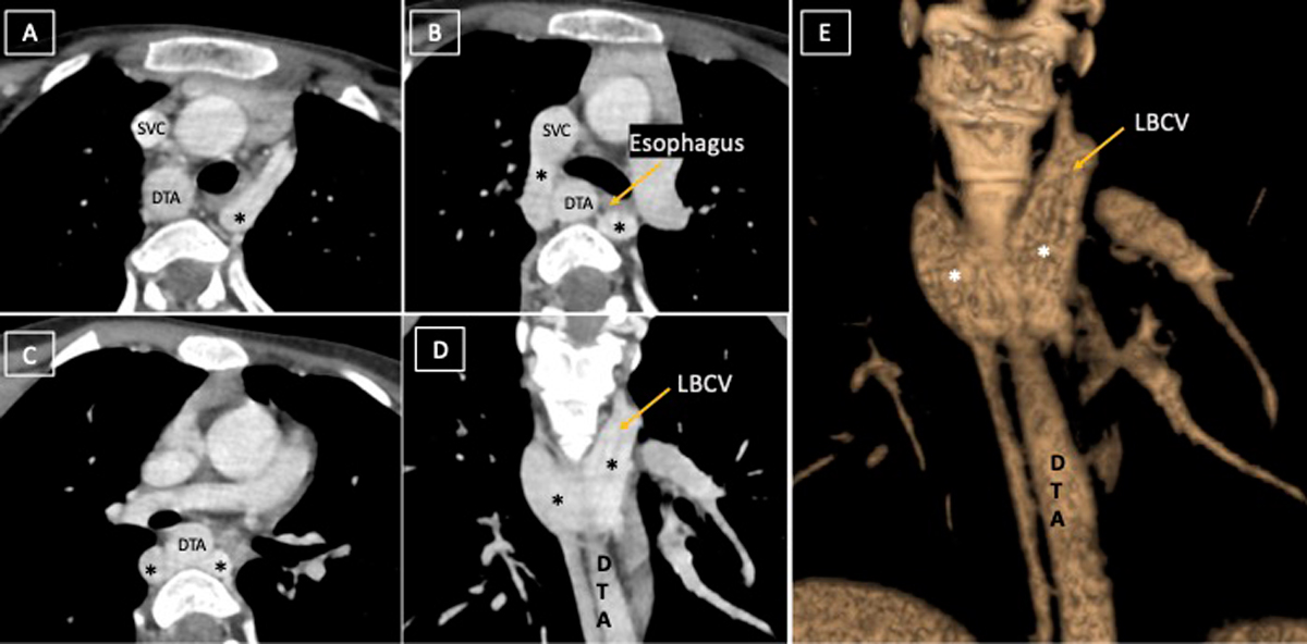
(A-C) Computed tomography angiography axial and (D) coronal image depicts retroesophageal U-shaped course of left brachiocephalic vein (LBCV, marked by *), which is joining the SVC. (E) Volume-rendered image depicts the anomalous course of LBCV. DTA: descending thoracic aorta; SVC: superior vena cava

DOI: https://doi.org/10.14797/mdcvj.1424 | Journal eISSN: 1947-6108
Language: English
Page range: 128 - 130
Submitted on: May 22, 2024
Accepted on: Sep 23, 2024
Published on: Nov 5, 2024
Published by: Houston Methodist DeBakey Heart & Vascular Center
In partnership with: Paradigm Publishing Services

© 2024 Archita Mahajan, Mansi Verma, Sushma Makhaik, published by Houston Methodist DeBakey Heart & Vascular Center
This work is licensed under the Creative Commons Attribution-NonCommercial 4.0 License.