
The normal course of the left brachiocephalic vein is anterosuperior to the aortic arch and its branches and then joins the right brachiocephalic vein to drain into the superior vena cava. An anomalous course of the brachiocephalic vein has an incidence of 0.2% to 1% in congenital heart diseases and is highest with tetralogy of Fallot (TOF).1 The left brachiocephalic vein coursing posterior to the esophagus is rare, with an incidence of approximately 0.19%.2 It is associated with cardiovascular malformations such as ventricular septal defect with or without pulmonary atresia, patent ductus arteriosus, and right-sided aortic arc.2 Genetic anomalies like 22q11 deletion are found to be associated with a retroesophageal course of the left brachiocephalic vein, hence genetic testing should be performed in such patients (Figure 1).
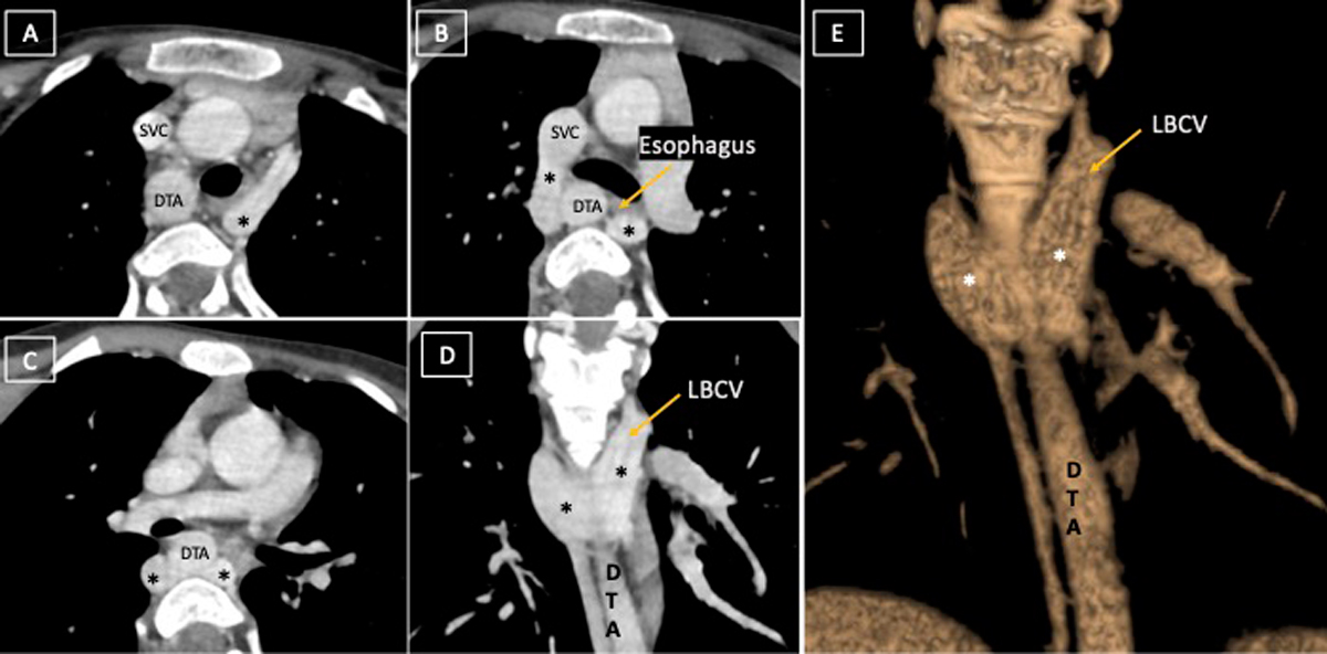
Figure 1
(A-C) Computed tomography angiography axial and (D) coronal image depicts retroesophageal U-shaped course of left brachiocephalic vein (LBCV, marked by *), which is joining the SVC. (E) Volume-rendered image depicts the anomalous course of LBCV. DTA: descending thoracic aorta; SVC: superior vena cava
The anomalous course is proposed to be present because of the persistent connection between the accessory hemizygous and left superior intercostal vein, which is due to interruption of the superior capillary plexus between the right and left anterior cardinal veins with failure of development of alternative inferior capillary plexus.2 The identification of the anomalous course of the brachiocephalic vein is clinically important because, for radiologists, the anomalous segment may be mistaken for an enlarged lymph node,3 an elevated right pulmonary artery in case of hypoplastic pulmonary artery, or an early branching right lobe pulmonary artery.4 For cardiologists, the left arm approach for central venous catheter and transvenous pacemaker insertion becomes difficult because, during cardiopulmonary bypass surgery, it may be seen as a persistent left superior vena cava (SVC), which further implicates establishment of alternate venous drainage.4 It may obscure the surgical field during construction of the subclavian-to-pulmonary-arterial shunt and may be confused with the pulmonary artery during right ventricular outflow tract reconstruction. Cannulation of the SVC with an anomalous brachiocephalic vein must be done with care to avoid obstruction of the anomalous vein. Its accidental manipulation during posterior mediastinal surgeries can lead to intraoperative bleeding.2
Competing Interests
The authors have no competing interests to declare. A written informed consent was obtained from the parent of the patient.