
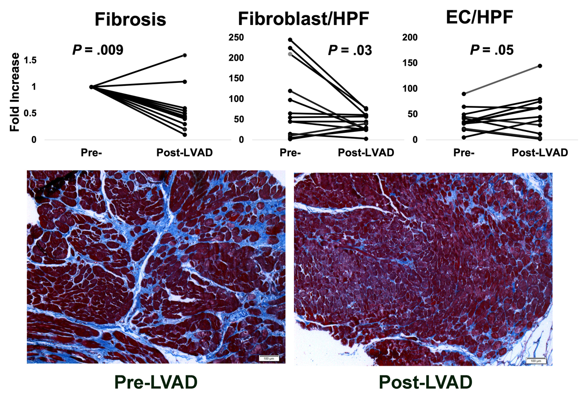
Figure 1
Left ventricular tissue from heart failure patients supported by LVAD demonstrates a decrease in fibrosis compared to that seen in their pre-LVAD sample (paired patient samples used). This decrease is accompanied by a reduction in fibroblast cell count per HPF and a concomitant increase in EC number. The decrease in fibrosis (blue) is visually evident using Masson’s Trichrome staining. LVAD: left ventricular assist device; HPF: high-power field; EC: endothelial cells (original figure)
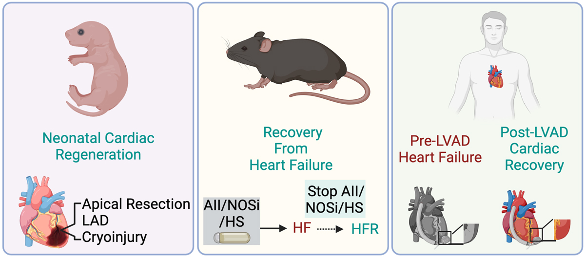
Figure 2
Models of cardiovascular regeneration and recovery. Neonatal mice have the intrinsic ability to regenerate the heart within 1 week of birth. Apical resection of LAD artery ligation and cryoprobe are used to create cardiac injuries. Depending on the methods and timing of the injury, the neonatal heart will regenerate to a variable degree, with some amount of fibrosis. In adult mice, heart failure is induced by the administration of angiotensin II, L-NAME (NOS inhibitor), and HS water. After cessation of these agents, cardiac chamber size and function recover, along with increased vascularization and reduced fibrosis. In humans, LVAD implantation facilitates heart recovery, and the paired tissue obtained from LVAD implantation (pre-LVAD) and from the native heart at the time of transplantation (post-LVAD) are useful for studying the cellular and molecular events during heart failure recovery. Image created with BioRender.com. LAD: left anterior descending; NOS: nitric oxide synthase; HS: high salt; LVAD: left ventricular assist device; AII: angiotensin II; NOSi: NOS inhibitor; HFR: recovery from heart failure