
Introduction
A fat embolism (FE) is defined as the presence of fat droplets within the peripheral circulation, and it often occurs after trauma. It was first described in humans in 1873 by Von Bergman.1 Fat embolism can lead to a systemic clinical presentation referred to as FE syndrome (FES), a multisystem disease affecting the lungs, brain, and skin and leading to respiratory insufficiency, neurological deficits, and petechiae, respectively.2 It is not often diagnosed because it is highly challenging to detect an FE in transit. Furthermore, the evaluation of FES remains a challenge for most clinicians due to its symptoms overlapping with many other conditions, such as pulmonary embolism, meningitis, or thrombotic thrombocytopenic purpura.
Here, we present a rare case of a patient in whom fat microemboli were detected and diagnosed during a spectral Doppler ultrasound. No signs or symptoms of FES (ie, skin rashes, respiratory or neurological complications) were observed in the patient.
Case
A 77-year-old female sought care at the emergency department with chief complaints of transient loss of consciousness and arm pain. After the episode, the patient presented with dyspnea and acute speech impairment. No motor weakness was present. The patient’s past medical history included hypertension, insulin-dependent diabetes mellitus, hypothyroidism, osteoporosis, vertebral compression fractures, and kyphoplasty of T12, L1, and L2 (postoperative day 6).
On physical examination, the patient was afebrile, tachycardic, hypertensive, and normo-saturated. The patient described severe right shoulder pain, and she was evaluated for syncope versus non-syncope conditions. As part of the diagnostic work-up, a right arm x-ray and venous duplex ultrasound of the upper extremities and neck were ordered. X-ray results showed a comminuted and impacted fracture of the right humeral neck. The patient’s family maintained that she possibly sustained her fracture while being lifted for transport by the emergency services. The patient was evaluated by orthopedics, who determined no acute intervention was needed and provided the patient with a sling brace.
The venous duplex ultrasound of upper extremities and neck suggested no evidence of venous thrombosis; however, during the spectral Doppler, multiple abnormal signals were detected in the right subclavian vein. These traces were intermittent and short, commonly referred to as high-intensity transient signals (HITS), indicative of an FE (Figure 1). On the right side, the brachial vein, cephalic vein, and basilic vein were not visualized due to the presence of the sling brace. However, the right internal jugular vein was clearly visualized, showing normal blood flow and no evidence of HITS. Examination of the left veins also reveals an absence of HITS. Had artificial interference been present, it would have been observed on both sides. Specifically, the left subclavian vein exhibited normal flow and compressibility, and no HITS were detected, as shown in Figure 2. The patient did not develop pulmonary or neurological symptoms. She was diagnosed with a urinary tract infection as the most likely cause of the transient loss of consciousness due to metabolic disturbance.
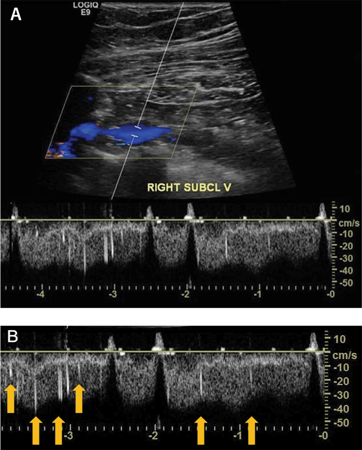
Figure 1
Venous duplex ultrasound of the right subclavian vein. (A) The rhomboid box illustrates the color Doppler showing blue blood flow without any visible clot. (B) Spectral Doppler (Zoom) shows multiple high intensity transient signals representing fat microemboli (denoted by yellow arrows).
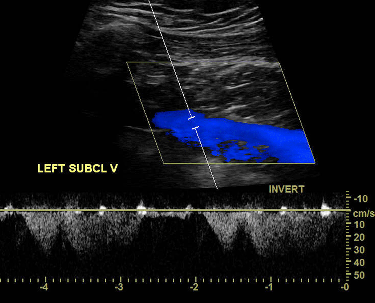
Figure 2
Venous duplex ultrasound of the left subclavian vein. Doppler image demonstrates normal blood flow and no detection of high intensity transient signals in the spectral Doppler.
Discussion
The reported incidence of FE is variable and likely inaccurate because it is difficult to diagnose and frequently presents in subclinical ways. The cause of FE may be traumatic or nontraumatic. Traumatic causes are commonly fractures, postoperative orthopedic procedures, and fat grafting.3 Nontraumatic causes include hemoglobinopathies and fatty liver, among others.
There are two main theories of FE pathogenesis.3 The first theory is mechanical and proposes that bone marrow manipulation releases fat droplets into the venous circulation. The second theory is biochemical and postulates that hormonal changes cause the release of free fatty acids, leading to inflammatory effects.4 Fat embolization is usually diagnosed in the context of FES, or through autopsy findings or macroscopic visualization during procedures. A diagnosis of FES is made via clinical and laboratory criteria but remains challenging.5
Although FE is a well-recognized phenomenon, it is rarely detected or reported. In the literature, we found just one case report of FE detected through HITS in spectral Doppler of the femoral vein. This finding was observed in a patient who underwent total knee arthroplasty.6 On grayscale ultrasound, FE has been described as the presence of a focal hyperechoic mass within the lumen of the vein, with normal compressibility and flow. In transesophageal echocardiography and transcranial Doppler (TCD), it is detected by the presence of HITS.7
Here, we present the first case of HITS detected in the upper extremities during spectral Doppler. HITS were first described in TCD as abnormal, high-intensity signals. These signals were attributed to turbulent blood flow or air bubble microemboli.8 Later, HITS were differentiated from artifact signals and attributed to formed-element emboli (fat or platelet-fibrinogen emboli) and bubble microemboli, suggesting that TCD could be a useful technique to diagnose cerebral embolism.9
In our case, the patient’s venous ultrasound had these HITS present only in the right subclavian vein; no HITS were found in the right axillary or internal jugular veins. This result suggests that the origin of the FE could be the proximal humeral fracture. The patient also underwent recent kyphoplasty, creating a second possible source. However, we note that HITS were visualized in a distal location of the subclavian vein, and the humeral fracture was much more recent than the kyphoplasty, making the latter source less likely.
One critical component of this interpretation is the ability to differentiate between true embolic signals and artifact signals. Embolic signals are transient, sharp, of short duration, have higher frequency intensity, and unidirectionality within the spectrum. In contrast, artifact signals tend to be bidirectional, with high intensity at low frequencies.10 Here, no clot was observed in the color Doppler, but HITS were detected in the spectral Doppler, suspected to be due to fat microembolization. These signals are identified based on our extensive experience with HITS in TCD. Previous studies have shown that fat emboli produce distinct spectral, which trained clinicians can reliably identify.7, 10
One important limitation of our report is the lack of saved sound in the duplex venous ultrasound. Typically, microembolic signals produce an audible “chirp,” but these sounds were not recorded due to oversight by the sonographer technician. It is important to acknowledge that sound is a valuable criterion for microembolic signals but not definitive for diagnosis.11
Conclusion
In conclusion, FES is known for occurring after musculoskeletal trauma and postsurgical procedures, yet it remains challenging to detect. FES treatment is based on early diagnosis and supportive therapies. Close pulmonary and neurological monitoring is indicated in positive diagnoses of FES. In high-risk populations, clinicians should be aware that FE can be detected as HITS in spectral Doppler. Although not every FE progresses to FES, FES can be a potentially fatal disease, and the variety of clinical symptoms that occurs can easily be mistaken for other diseases.12 Early diagnosis of FE can be crucial when clinicians suspect FES, allowing for rapid treatment and reduced risk of serious complications.
Acknowledgements
We would like to express our gratitude to our medical writer, Michelle C. Swick, PhD, ELS, for her valuable edits.
Competing Interests
The authors have no competing interests to declare.