
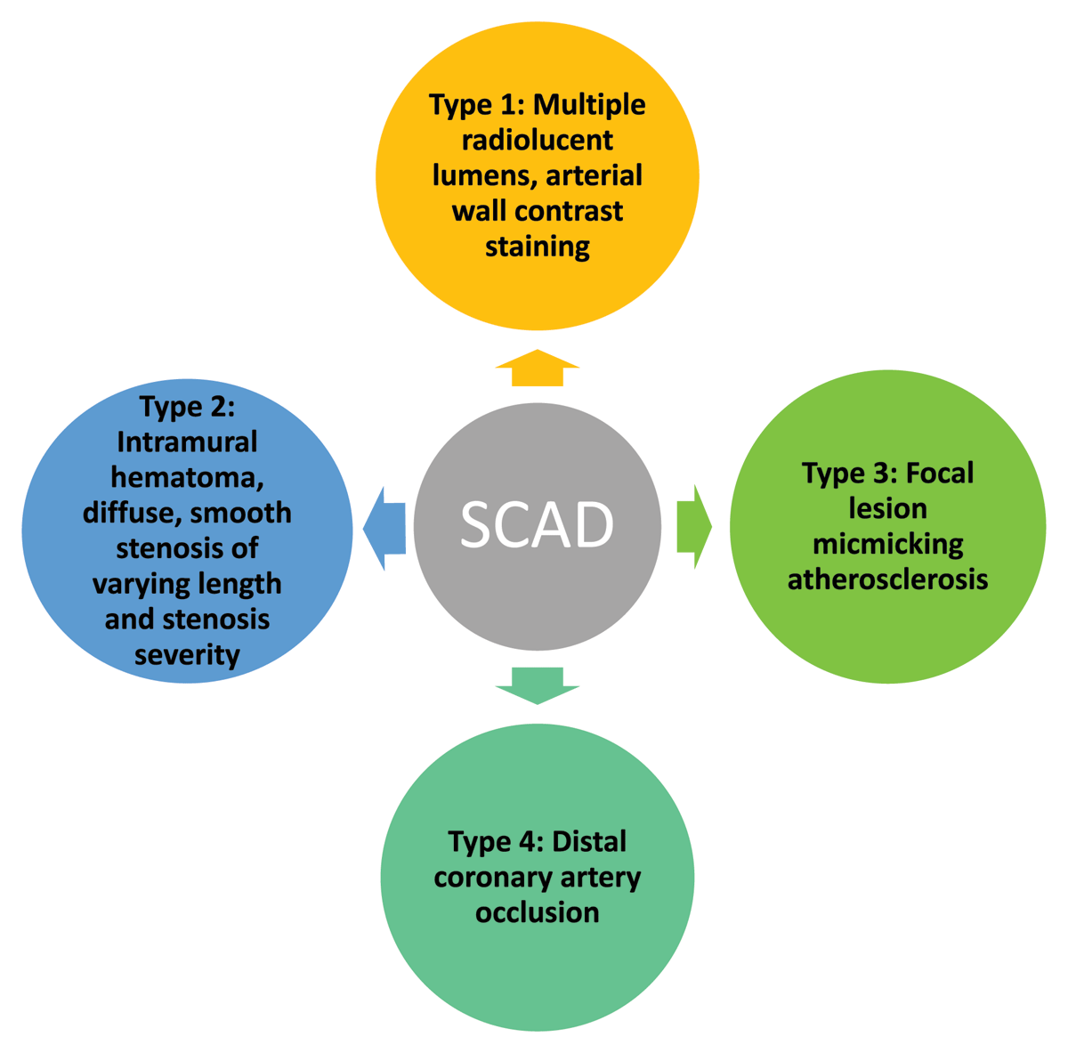
Figure 1
Yip-Saw angiographic classification for spontaneous coronary artery dissection (SCAD).
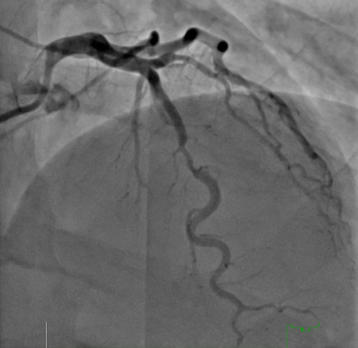
Figure 2
Invasive coronary angiogram of type 3 spontaneous coronary artery dissection (SCAD) in a 54-year-old woman presenting with non-ST-elevation myocardial infarction (NSTEMI). Type 3 SCAD is the most difficult type to distinguish from obstructive coronary artery disease.
Table 1
American Heart Association, American College of Cardiology, and European Society of Cardiology guidelines for the management of patients with chronic coronary artery disease. COVADIS: Coronary Vasomotor Disorders International Study Group; CMD: coronary microvascular disease; CAD: coronary artery disease; FFR: fractional flow reserve; CTA: computed tomography angiography; ECG: electrocardiography; IMR: microcirculatory resistance; TIMI: Thrombolysis in Myocardial Infarction
| COVADIS DIAGNOSTIC CRITERIA FOR CMD |
|---|
| Symptoms of myocardial ischemia |
|
| Absence of obstructive CAD (< 50% diameter reduction or FFR > 0.80) by |
|
| Objective evidence of myocardial ischemia |
|
| Evidence of impaired coronary microvascular function |
|
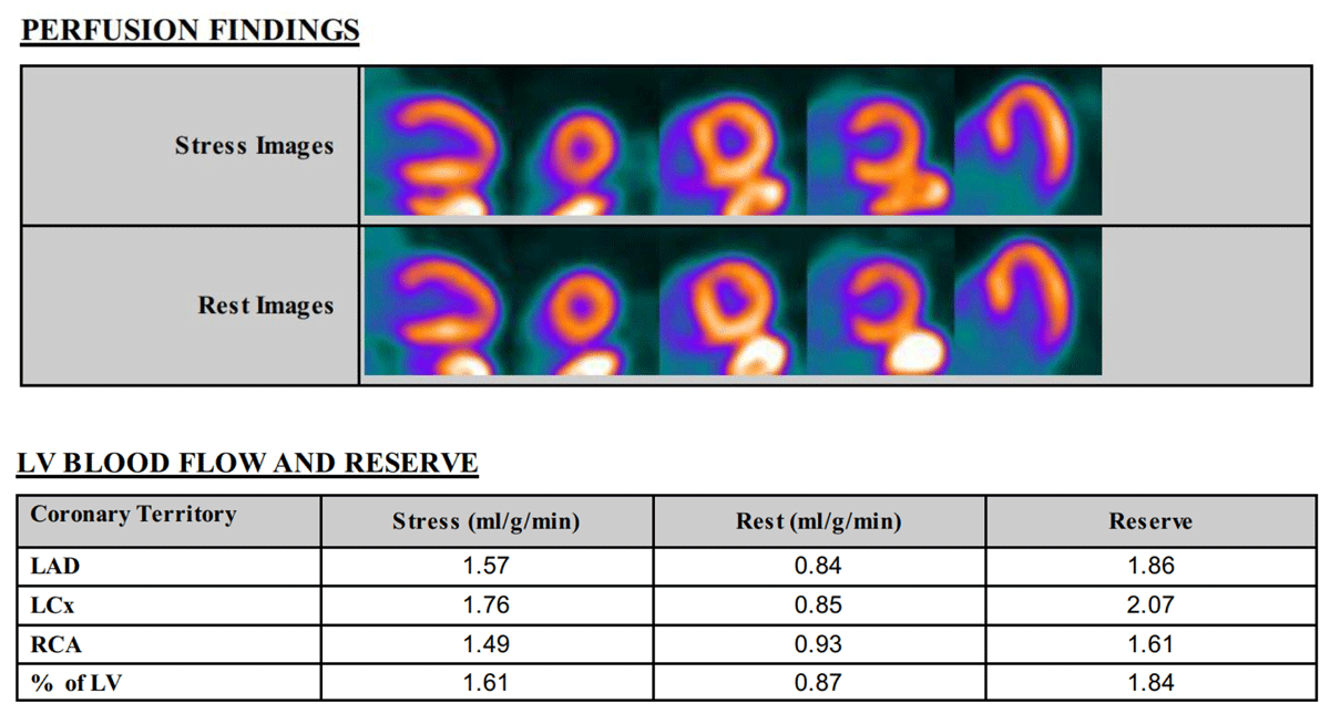
Figure 3
Positron emission tomography with myocardial flow reserve (MFR) showing normal perfusion with impaired MFR of 1.84 (normal MFR > 2) in a 76-year-old woman with microvascular angina. LV: left ventricular; LAD: left anterior descending artery; LCx: left circumflex artery; RCA: right coronary artery
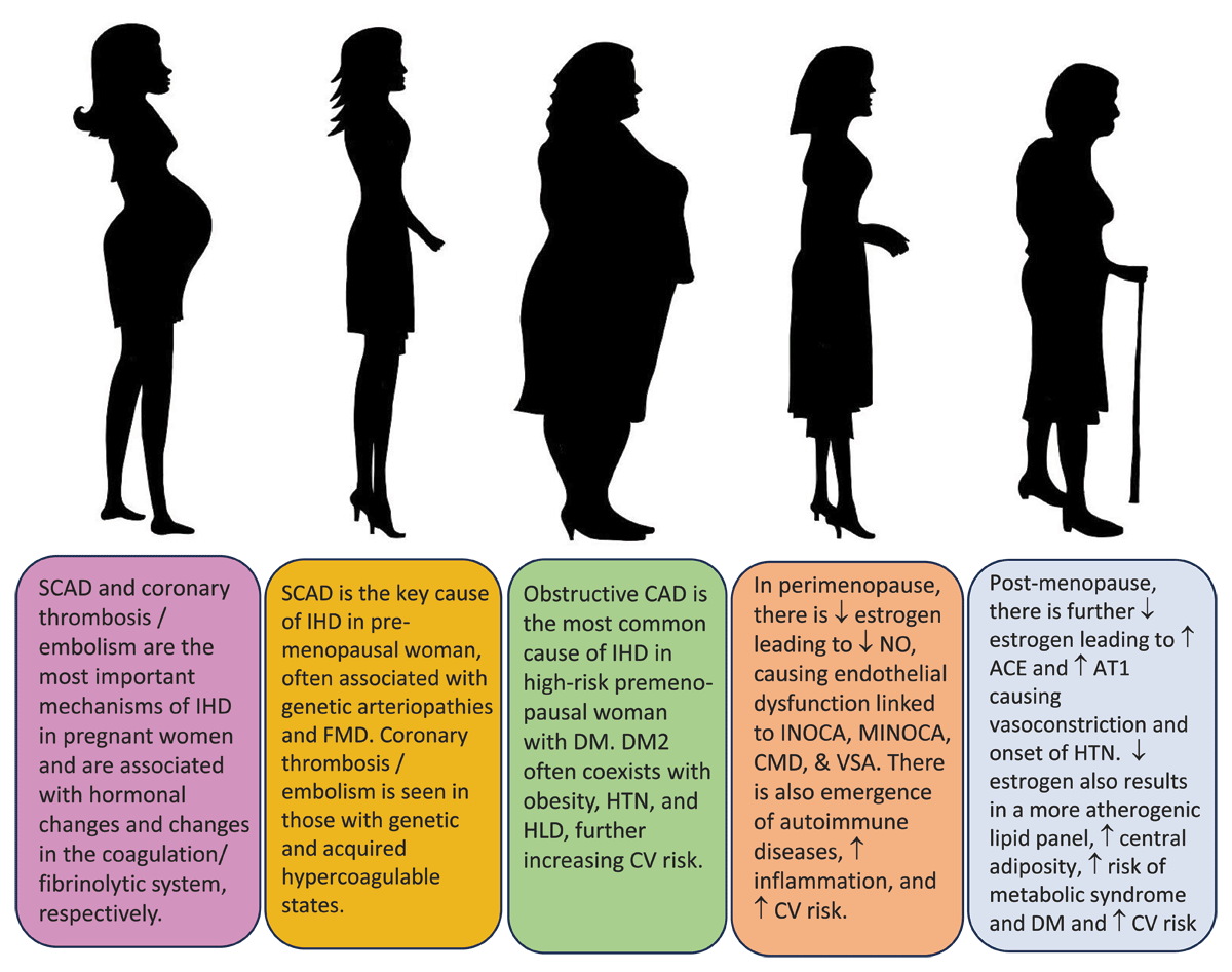
Figure 4
Spectrum of ischemic heart disease (IHD) in women. SCAD: spontaneous coronary artery dissection; FMD: fibromuscular dysplasia; DM2: diabetes mellitus type 2; CAD: coronary artery disease; CV: cardiovascular; CMD: coronary microvascular dysfunction; INOCA: ischemia with no obstructive coronary arteries; MINOCA: myocardial infarction with no obstructive coronary arteries; VSA: vasospastic angina; ACE: angiotensin converting enzyme; AT1: angiotensin 1 receptor; HTN: hypertension