
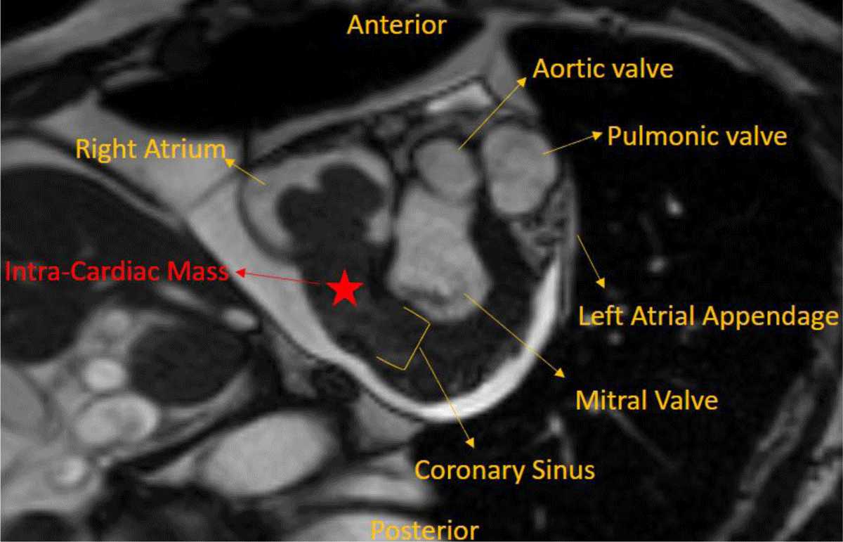
Figure 1
Left atrial view showing the large mass in the right atrium extending into the coronary sinus.
Video 1
Axial plane cardiac magnetic resonance image demonstrating the extent of the intracardiac mass, which starts in the right atrium and extends into the coronary sinus. Incidentally, the patient has a large pericardial effusion without tamponade physiology. See also at https://youtu.be/y9w65OyMKNs.
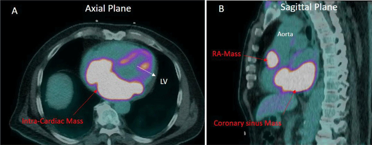
Figure 2
(A) Axial plane image demonstrating intense fluorodeoxyglucose (FDG) uptake in the intracardiac mass (SUV Max 18). (B) Sagittal plane image demonstrating a second intracardiac mass in the wall of the right atrium, which also has intense FDG uptake (SUV Max 12). Intense FDG uptake in both masses is consistent with a malignant tumor. LV: left ventricle; RA: right atrium
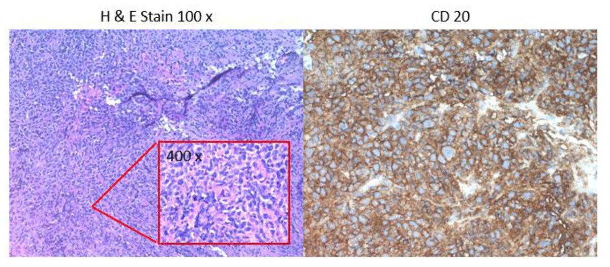
Figure 3
Histologic sections of the atrial mass biopsy showed a diffuse infiltrate of large atypical lymphocytes with areas of necrosis. Immunohistochemical stains showed that the lymphocytes were positive for CD20, BCL6, and MUM-1 and negative for CD10. FISH studies were negative for rearrangements of MYC, BCL2, and BCL6. The overall findings were those of diffuse large B-cell lymphoma, non-germinal center immunophenotype. FISH: fluorescence in situ hybridization