
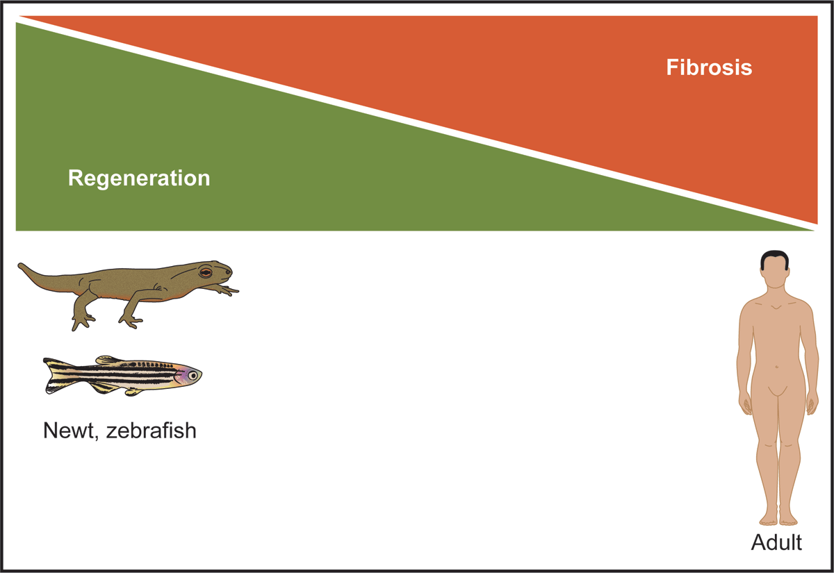
Figure 1
Lower organisms have a remarkable regenerative capacity following myocardial injury. The adult newt and zebrafish are examples of lower organisms that can completely regenerate their hearts following an acute injury. This regenerative process is due to cardiomyocyte dedifferentiation and proliferation in the absence of a fibroproliferative (ie, scar) response. In contrast, the adult mammalian heart has a very limited regenerative capacity, and repair of the adult heart is associated with extensive fibrosis in order to stabilize the injured heart. Figure is adapted from references 17-20.
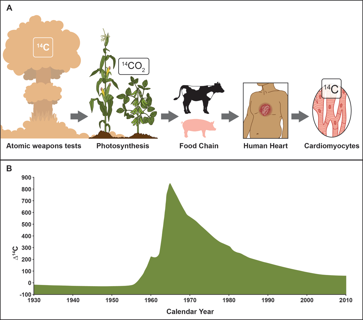
Figure 2
The adult human heart has a low turnover of cardiomyocytes. (A) Radiocarbon dating of the human heart relies upon a pulse (B) of 14C via the aboveground atomic tests, which was then incorporated into vegetation (via photosynthesis) and consumed by animals and humans. The levels of 14C found in cardiomyocyte nuclei were used to determine the birth date of the cardiomyocytes. These studies support the notion that 0.5% to 1.0% of human cardiomyocytes turn over each year. While this number is relatively low, these studies emphasize that pathways and subpopulations of cells are present and could be enhanced as a therapeutic initiative. Figure is adapted from references 36–38.
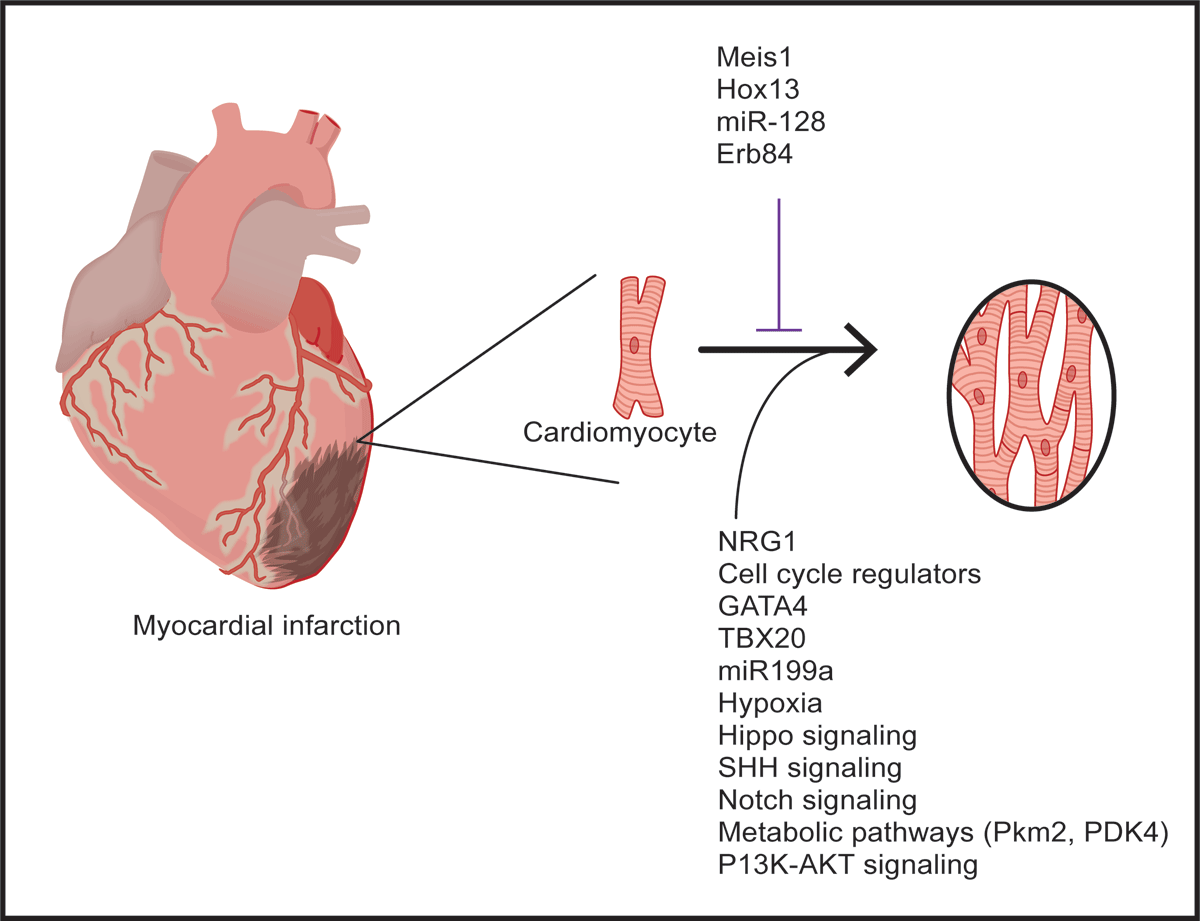
Figure 3
Factors and pathways promote cardiomyocyte proliferation in the adult mammalian heart. Schematics outlining several of the factors, environmental conditions, and pathways that have been reported to promote cardiomyocyte proliferation following injury of the adult mammalian heart. Figure is adapted from references 36-38.