
Table 1
Causes of restrictive cardiomyopathy.
| Infiltrative (Accumulation of substance between myocytes) | Amyloidosis (inherited/acquired) Hurler’s disease (inherited) Hunter’s disease (inherited) Gaucher’s disease (inherited) Sarcoidosis (acquired) |
| Storage disorders (Accumulation of substance within myocytes) | Fabry’s disease (inherited) Glycogen storage disease (inherited) Iron overload cardiomyopathy (inherited/acquired) |
| Non-infiltrative | Scleroderma (acquired) Idiopathic restrictive cardiomyopathy (acquired/inherited) Pseudoxanthoma elasticum (inherited) |
| Endomyocardial | Endomyocardial fibrosis (multifactorial) Hypereosinophilic syndrome (acquired) Endocardial fibroelastosis (inherited) Carcinoid syndrome (acquired) Drug induced: hydroxychloroquine, ergotamine, methysergide (acquired) Radiation-induced cardiomyopathy (acquired) |
Table 2
Diagnostic features of primary and secondary RCM. LV: left ventricular; IOC: iron overload cardiomyopathy; HFE: hereditary hemochromatosis; LGE: late gadolinium enhancement; alpha-Gal A: alpha-galactosidase A; GI: gastrointestinal; AV: atrioventricular; RCM: restricted cardiomyopathy
| TYPE | CLINICAL FINDINGS | LABORATORY INVESTIGATION/GENETIC TESTING | ECHOCARDIOGRAPHY | CARDIAC MAGNETIC RESONANCE IMAGING | ENDOMYOCARDIAL BIOPSY | TREATMENT |
|---|---|---|---|---|---|---|
| Idiopathic RCM | Any age group, family hx +/-, skeletal myopathy +/- | Genetic testing: mutations in sarcomere encoding proteins/desmin | +/- increased LV wall thickness | Myocyte hypertrophy, disarray, and interstitial fibrosis | Symptomatic treatment; advanced heart failure therapies | |
| Iron overload cardiomyopathy | Primary: skin pigmentation, diabetes, liver dysfunction Secondary IOC: underlying hematological disorder | Elevated serum ferritin and transferrin saturation level Primary IOC: HFE gene mutation | +/- increased LV wall thickness Dilated cardiomyopathy at later stages | Decreased T2* relaxion time | Prussian blue staining+ | Phlebotomy, iron chelators |
| Fabry’s disease | Cardiac manifestation: 30s, later in females | Absent or reduced leukocyte alpha-Gal A activity Genetic testing: gene encoding alpha-Gal A | Increased LV wall thickness | Midmyocardial LGE pattern in basal inferolateral wall | Concentric lamellar bodies in the sarcoplasm of myocytes | L-algalsidase beta |
| Endomyocardial fibrosis | Tropical countries, endemic disease, malnutrition Bimodal peak at 10 and 30 years | Eosinophilia +/- | Mural thrombus in apical and valvular pockets Obliteration and retraction of ventricles, endomyocardial thickening, AV valve regurgitation | Subendocardial diffuse LGE pattern from apex to valvular region, thrombus +/- | Fibrosis, +/- eosinophilic infiltration | Steroids in early phase, endomyocardial resection with valve repair or replacement |
| Hypereosinophilic syndromes | Skin rash, pulmonary, GI, neurological manifestations > temperate zones | Eosinophilia | Endomyocardial thickening, AV valve regurgitation, mural thrombus | Subendocardial patchy or diffuse LGE pattern, high intensity on T2-WI, +/- thrombus | Eosinophilic infiltrates, fibrosis | Steroids +/- Hydroxyurea/interferon alfa Imatinib: F1P1L1-PDGFRA mutation Endomyo-cardectomy with valve repair or replacement |
| Drug-induced RCM | Hydroxychloroquine | Increased wall thickness | Curvilinear bodies, lysosomes, myeloid bodies, glycogen granules and myocyte vacuolation seen on electron microscopy | Withdrawal of the drug | ||
| Radiation-induced RCM | Mediastinal radiation, latent period 10-15 years | Valvular calcification | Transmural or subendocardial LGE, perfusion defects + | Fibrosis | Symptomatic treatment |
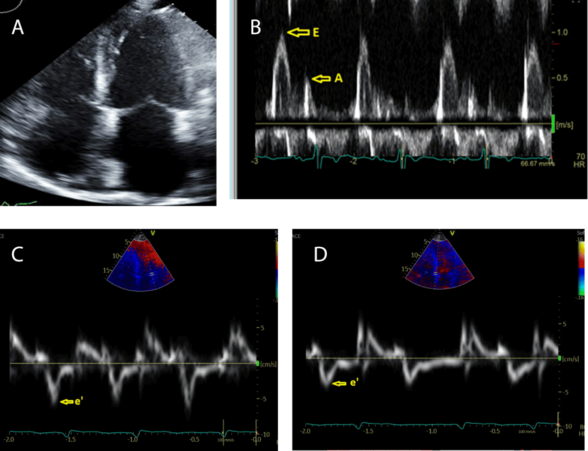
Figure 1
(A) Apical 4-chamber view showing biatrial enlargement. (B) Mitral inflow doppler showing increased E/A ratio (feature of diastolic dysfunction). (C, D) Lateral and septal mitral annular tissue doppler showing decreased e’ velocity.
Table 3
Echocardiography findings in restrictive cardiomyopathy. EF: ejection fraction; LV: left ventricular.
| Normal or mildly reduced EF |
| Normal or slightly increased LV wall thickness |
| Normal or slightly decreased LV cavity |
| Biatrial enlargement |
Diastolic dysfunction
|
Table 4
Iron overload cardiomyopathy (IOC).
| Etiology | Primary IOC | Hereditary hemochromatosis |
| Secondary IOC | Hereditary anemia
Chronic liver disease Increased dietary intake | |
| Diagnostic evaluation | Biomarkers | Serum ferritin > 200 ng/mL in premenopausal women or > 300 ng/mL in men and postmenopausal women Serum transferrin > 45% in men and > 55% in women |
| Echocardiography | Early stage: diastolic dysfunction with pseudonormalization or restrictive filling pattern with or without atrial enlargement. Later stage: left and right cardiac chamber dilation with reduced left ventricular ejection fraction | |
| Cardiac magnetic resonance | T2* valve < 20 ms indicates IOC, < 10 ms indicates severe iron overload and poor outcomes |
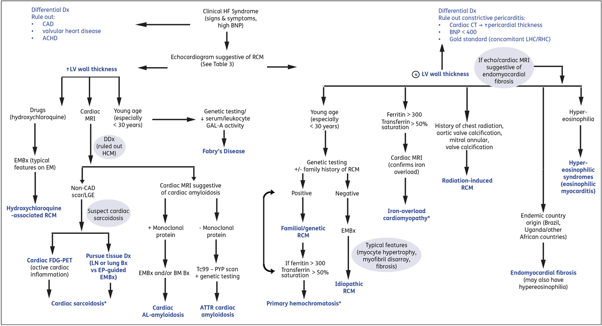
Figure 2
Suggested algorithm for evaluation of RCM. Please note that as RCM is a heterogenous disease process, there may be variability in clinical presentation (eg, cardiac hemochromatosis or sarcoidosis may also present with dilated phenotype and normal wall thickness).* This suggested approach is intended to assist with comprehension. Blue font indicates final diagnosis. Dx: diagnosis; DDx: differential diagnosis; CAD: coronary artery disease; HF: heart failure; ACHD: adult congenital heart disease; BM: bone marrow; EMBx: endomyocardial biopsy; RCM: restricted cardiomyopathy; MRI: magnetic resonance imaging; FDG-PET: fluorodeoxyglucose positron emission tomography; LV: left ventricular; Bx: biopsy; AL: light chain amyloidosis; ATTR: transthyretin amyloidosis; LGE: late gadolinium enhancement; Tc99-PYP scan: technetium-99 m pyrophosphate scintigraphy; AV: atrioventricular; GAL-A: alpha-galactosidase A; EP: electrophysiologic; LHC/RHC: left/right heart catheterization
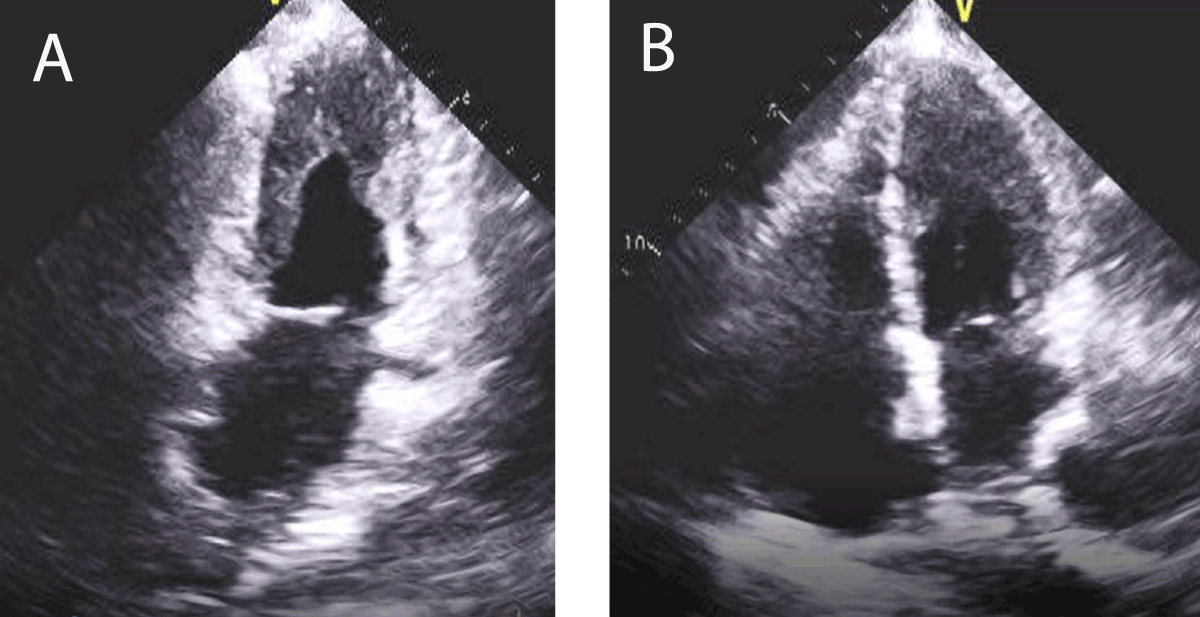
Figure 3
Apical 2-chamber and 4-chamber echocardiographic view showing biatrial enlargement, apical obliteration by thrombus.
Table 5
Hypereosinophilic syndromes. vWF: von Willebrand factor; AV: atrioventricular
| TYPE OF SYNDROME | DESCRIPTION |
|---|---|
| Hypereosinophilic syndromes | Absolute eosinophil count > 1.5 × 10⁹/L for longer than 1 month and eosinophil-mediated end organ damage |
| Primary Secondary Idiopathic | Stem cell and myeloproliferative disorders Parasitic, fungal infections, allergic conditions, solid tumors |
| Cardiac involvement | Acute necrotic phase: eosinophilic infiltration, degranulation, inflammation and necrosis Thrombotic phase: activation of vWF and tissue factor on damaged endocardium and thrombus formation Late fibrotic phase: recurrent inflammation leading to fibrosis |
| Echocardiography | Acute stage: no changes unless fulminant myocarditis Thrombotic/fibrotic stage: endomyocardial thickening/fibrosis, restrictive filling pattern, AV valve regurgitation due to fibrotic involvement of the valve apparatus |
| Treatment | Treat the underlying condition Acute phase: Corticosteroids +/- hydroxyurea/interferon alfa Latent stage: heart failure management Endomyocardectomy, valve repair or replacement |