
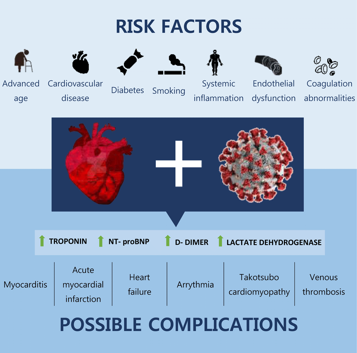
Figure 1
Risk factors for cardiovascular involvement of COVID-19 and possible complications.
Table 1
Summary of cardiac injury prevalence (defined as troponin elevation) in COVID-19 studies.12,18,19,20,21,22,23,24,25,26 ICU: intensive care unit.
| AUTHOR | N | (%) WITH ELEVATED TROPONIN | ASSAY USED | PATIENT SETTING |
|---|---|---|---|---|
| Metkus et al.20 | 243 | 51 | Troponin I or T | ICU |
| Giustino et al.21 | 305 | 62 | Troponin T | Inpatient |
| Huang et al.22 | 41 | 12 | Hypersensitive troponin I | Inpatient |
| Han et al.23 | 273 | 5.05% (outpatients) 23.33% (inpatients) 20% (ICU) | Hypersensitive troponin I | Outpatient, inpatient, ICU |
| Richardson et al.12 | 5,700 | 22.6 | Variety of assays | Inpatient |
| Petrilli et al.24 | 4,103 | 11.7 | Not reported | Outpatient and inpatient |
| Wang et al.25 | 138 | 7.2 | Troponin I | Inpatient |
| Zhou et al.26 | 191 | 17 | High-sensitivity troponin I | Inpatient |
| Guo et al.18 | 187 | 27.8 | Troponin T | Inpatient |
| Shi et al.19 | 416 | 19.7 | High-sensitivity troponin I | Inpatient |
Table 2
Main findings of studies utilizing CMR in COVID-19 patients.35,36,37,38,39,40,41,42,43,44 ECV: extracellular volume fraction; CMR: cardiac magnetic resonance; LGE: late-gadolinium enhancement; hsTNT: high-sensitivity troponin T.
| FIRST AUTHOR, YEAR | COUNTRY | TYPE OF STUDY | NO. OF PATIENTS | RESULTS | PRIMARY END POINTS | CONCLUSION |
|---|---|---|---|---|---|---|
| Joy38 2021 | UK | Prospective blind study | 74 recovered patients with mild cases vs. 75 controls | No difference in end points between recovered patients and controls | Cardiac involvement 6 months after recovery from mild COVID-19 | Mild COVID-19 in healthy patients does not result in cardiovascular abnormalities. |
| Raman37 2021 | UK | Observational cohort study | 58 recovered patients from moderate-severe COVID-19 vs. 30 matched controls | • 26% elevated basal myocardial T1 • No statistical difference in T2 and ECV | Cardiac involvement 2-3 months after recovery from COVID-19 | Multiorgan inflammation persists after recovery from moderate-severe COVID-19. |
| Starekova39 2021 | USA | Case series | 145 competitive athletes recovering from mild-moderate COVID-19 who underwent CMR 15 days after diagnosis | 1.4% had CMR findings consistent with myocarditis | Prevalence of myocardial involvement in competitive athletes recovering from COVID-19 | There is low prevalence of myocarditis in this population. |
| Daniels40 2021 | USA | Case series | 1,597 competitive athletes recovering from COVID-19 | 2.3% had CMR-diagnosed myocarditis, clinical and subclinical | Prevalence of myocarditis in competitive athletes recovering from COVID-19 | CMR screening in athletes recovering from COVID-19 should be considered for safe return to play. |
| Martinez41 2021 | USA | Cross-sectional study | 789 professional athletes with COVID-19 infection, irrespective of symptoms | 0.6% had CMR findings suggesting inflammatory heart disease | Prevalence of detectable inflammatory heart disease in professional athletes with prior COVID-19 infection | Few cases of inflammatory heart disease have been detected; safe return to play has been achieved. |
| Kotecha42 2021 | UK | Retrospective study | 148 patients with severe COVID-19 requiring hospitalization | 54% had LGE: • 26% myocarditis • 22% ischemia • 6% dual pathology | Assess myocardial injury in hospitalized COVID-19 patients after recovery | During recovery from severe COVID-19, myocarditis-like injury can be detected. Its functional consequence is not clear. |
| Puntmann36 2020 | Germany | Prospective observational cohort study | 100 recovered patients vs. 107 controls | • Abnormal CMR findings in 78% of recovered COVID-19 patients • 73% raised myocardial native T1 • 60% raised myocardial native T2 • 32% LGE • 22% pericardial involvement | Cardiac involvement after recovery from COVID-19 | CMR revealed cardiac involvement and ongoing myocardial inflammation in recovered COVID-19 patients. |
| Huang35 2020 | China | Retrospective study | 26 recovered patients | • 58% abnormal CMR • 54% myocardial edema • 31% LGE | Cardiac involvement after recovery from COVID-19 | A proportion of recovered COVID-19 patients had cardiac involvement on CMR. |
| Rajpal43 2020 | USA | Case series | 26 competitive athletes with mild COVID-19 | • 15% CMR findings consistent with myocarditis • 46% LGE | Detect cardiac involvement through CMR in competitive athletes recovering from COVID-19 | CMR may help stratify athletes recovering from COVID-19 as to risk of myocarditis. |
| Knight44 2020 | UK | Cross-sectional study | 828 hospitalized patients positive for COVID-19 or with a clinical diagnosis | • 586 patients had elevated hsTNT • 51 underwent CMR: 69% of them had myocardial injury | Underlying cause of troponin elevation in COVID-19 infection | Myocardial injury detected by CMR is common in hospitalized COVID-19 patients. |
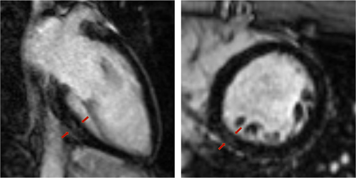
Figure 2
Cardiac magnetic resonance image of 25-year-old healthy male complaining of persistent chest pain and palpitations 5 weeks after COVID-19 diagnosis. Mild late-gadolinium enhancement can be visualized in the basal inferior and inferolateral myocardial wall. Holter monitoring showed frequent premature ventricular beats.