
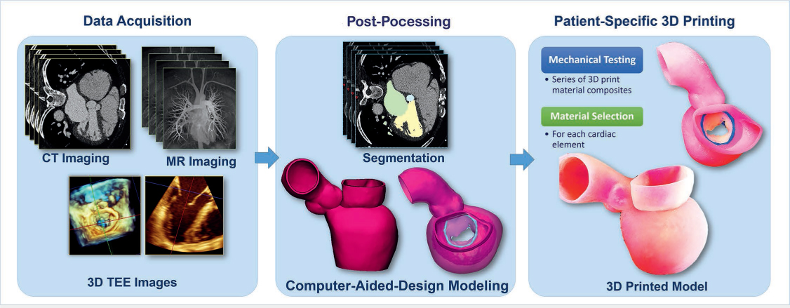
Figure 1.
3-dimensional (3D) printed model development workflow. Patient-specific 3D printed modeling starts with high-quality data acquisition from computed tomography (CT), magnetic resonance (MR) imaging, and 3D transesophageal echocardiography (TEE) images. The next step is imaging post-processing that includes geometry segmentation and computer-aided design modeling. The models are 3D printed using multiple materials that were previously selected based on mechanical testing.
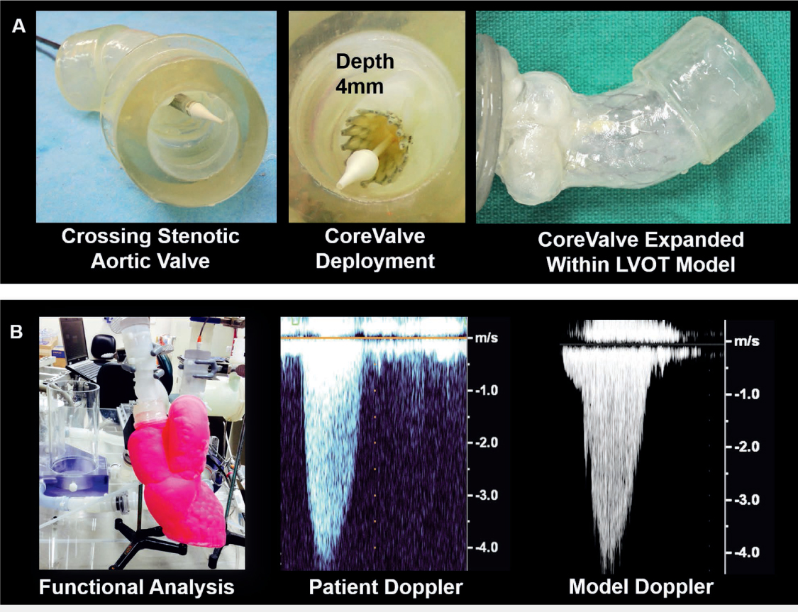
Figure 2.
Patient-specific 3-dimensional (3D) printed aortic stenosis modeling. (A) Patient-specific aortic stenosis with calcific structures used for benchtop implantation and sizing of the CoreValve device. (B) Patient-specific 3D printed aortic stenosis model with left ventricle, left atrium, and left ventricular outflow tract (LVOT) used for functional analysis and echocardiographic assessment within the flow loop under pressurized flow conditions (left panel). Color Doppler data acquired in the model within the flow loop (right panel) compared favorably with the color Doppler of the actual patient (middle panel). Images adapted with permission from Vukicevic et al.10
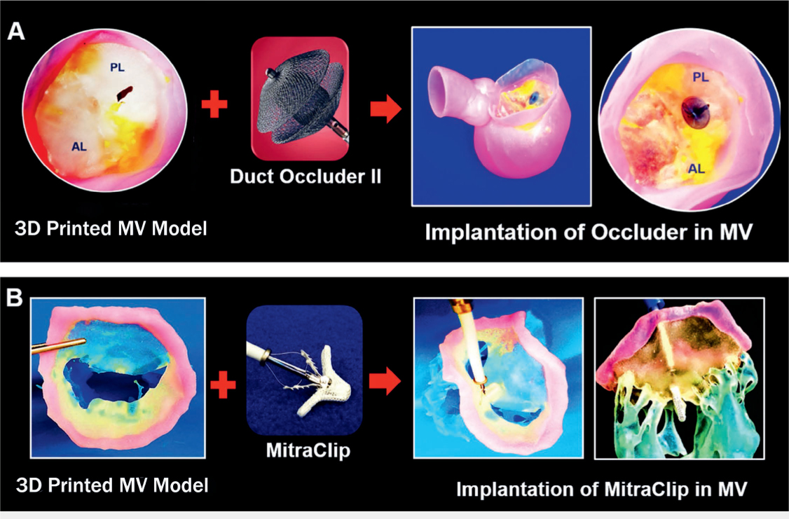
Figure 3.
Patient-specific 3-dimensional (3D) printed models used for structural heart interventions planning. (A) Procedural planning of mitral valve (MV) with perforated posterior leaflet; occluding device selection and sizing was performed using 3D printed patient-specific mitral valve model. (B) Benchtop implantation of MitraClip device (Abbott) within the patient-specific 3D printed mitral valve model with chordae and papillary muscles. Figures adapted with permission from Vukicevic et al.10 AL: anterior leaflet; PL: posterior leaflet
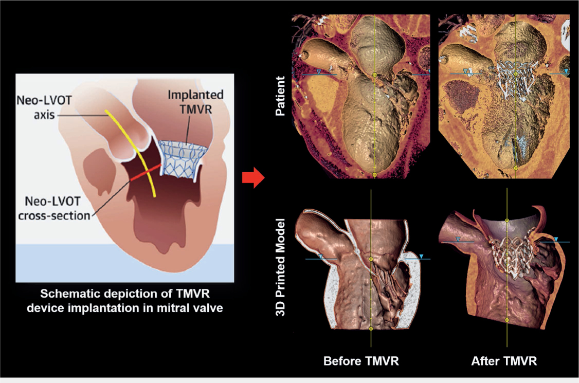
Figure 4.
Transcatheter mitral valve replacement (TMVR) procedure planning using 3-dimensional (3D) printed model. Schematic depiction of TMVR device deployment and evaluation of neo-left ventricular outflow tract (LVOT) cross-section area (left panel; adapted with permission from Vukicevic et al.)4 Computed tomography imaging of the patient before and after the TMVR procedure (upper panel) compared well with the CT scan of the 3D printed model before and after the TMVR device implantation (lower panel).
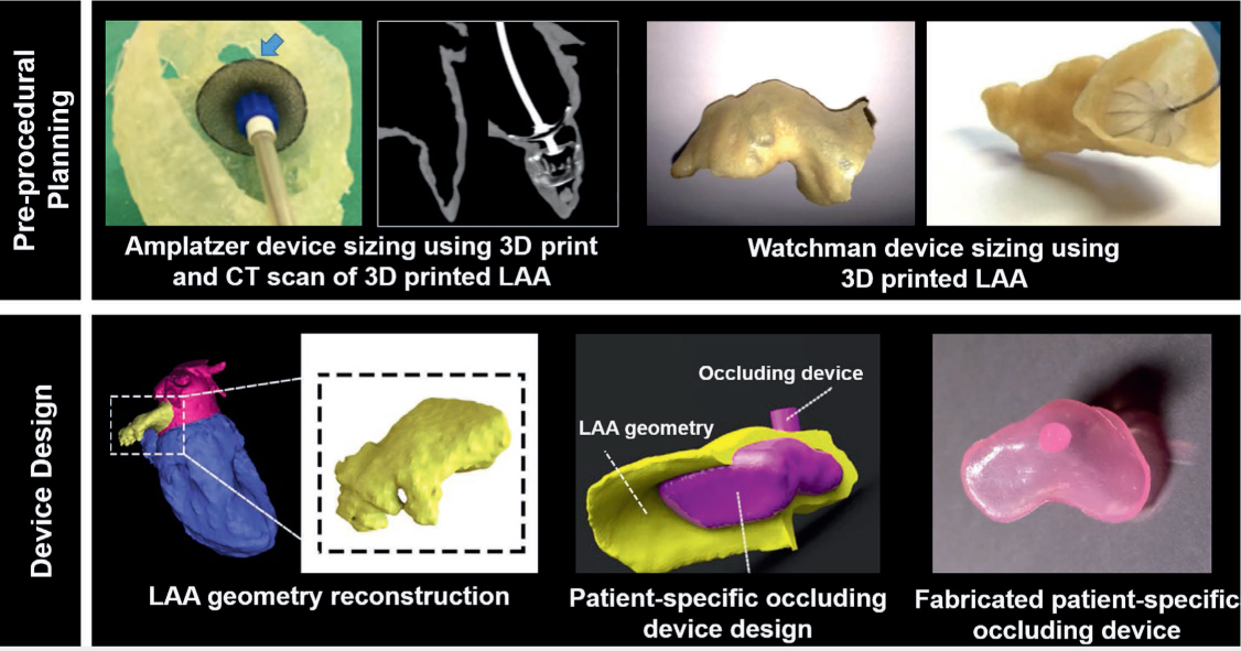
Figure 5.
Preprocedural planning and device design based on patient-specific 3-dimensional (3D) printed left atrial appendage (LAA) models. (Upper left panel) Benchtop implantation of 25 Amplatzer (Abbott) occluding device in 3D printed LAA shows that the occluding device, which was selected based on measurements from computed tomography (CT) and echocardiographic images, is oversized. A smaller 22 Amplatzer device (shown in CT image of the model) was eventually implanted in the patient. Upper left panel figures adapted with permission from Hachulla et al.18 (Upper right panel) CT-based 3D printed model of LAA was developed for planning the Watchman (Boston Scientific Corporation) device implantation. Upper right panel figures adapted with permission from Obasare et al.19 Device design based on the patient's geometry was developed using the 3D printed modeling. Bottom panel adapted with permission from Robinson et al.20
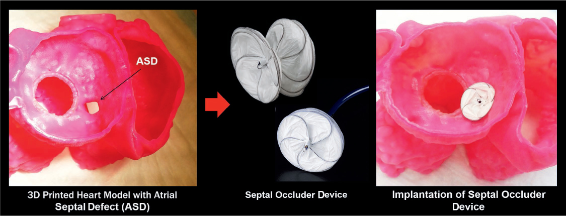
Figure 6.
Preprocedural planning of an atrial septal defect (ASD) was performed using a patient-specific heart model. Adapted with permission from Vukicevic et al.4