
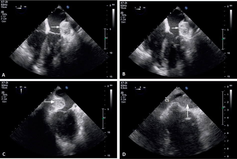
Figure 1.
Intraoperative transesophageal echocardiogram illustrating the left atrial intramural hematoma in the (A, B) midesophageal four-chamber view, (C) midesophageal two-chamber view, and (D) midesophageal coronary sinus view. The arrows indicate the hematoma. CS: coronary sinus. Videos are available on the journal website.