
Case Description
A 64-year-old gentleman with severe aortic stenosis was admitted for surgical aortic valve replacement. A bioprosthetic valve was implanted. Intraoperative transesophageal echocardiogram (TEE) immediately after cardiopulmonary bypass showed a well-seated bioprosthetic aortic valve with trivial intravalvular aortic insufficiency and no perivalvular leaks. The other cardiac valves showed normal function. Normal left ventricular (LV) and right ventricular (RV) systolic functions were noted, with no regional wall motion abnormalities.
The patient came off bypass uneventfully, and the chest was closed. At this time, TEE demonstrated interval development of a mass in the posterolateral part of the left atrium, adjacent to the left atrial appendage and left upper pulmonary vein and extending to the coronary sinus (Figure 1, see online videos 1–3). Consequently, the patient went back on cardiopulmonary bypass for further exploration, during which the surgical team discovered an intramural hematoma with no active hemorrhage. The hematoma was thought to be secondary to surgical trauma. TEE showed that valvular and ventricular function was unchanged.
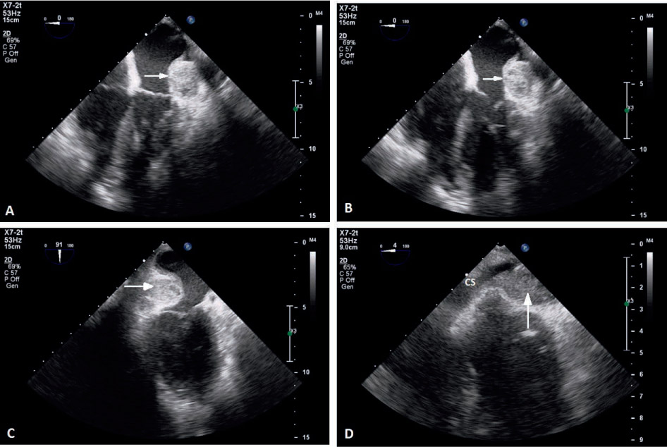
Figure 1.
Intraoperative transesophageal echocardiogram illustrating the left atrial intramural hematoma in the (A, B) midesophageal four-chamber view, (C) midesophageal two-chamber view, and (D) midesophageal coronary sinus view. The arrows indicate the hematoma. CS: coronary sinus. Videos are available on the journal website.
Reports of left atrial intramural hematomas as a complication of cardiac valve surgery are exceedingly rare. The patient exhibited stable hemodynamics, and a conservative approach was deemed appropriate with planned future reassessment by echocardiography. The patient had a benign perioperative course and was discharged home. He was doing well at 4-month follow up. Transthoracic echocardiography at that time showed resolution of the hematoma.