
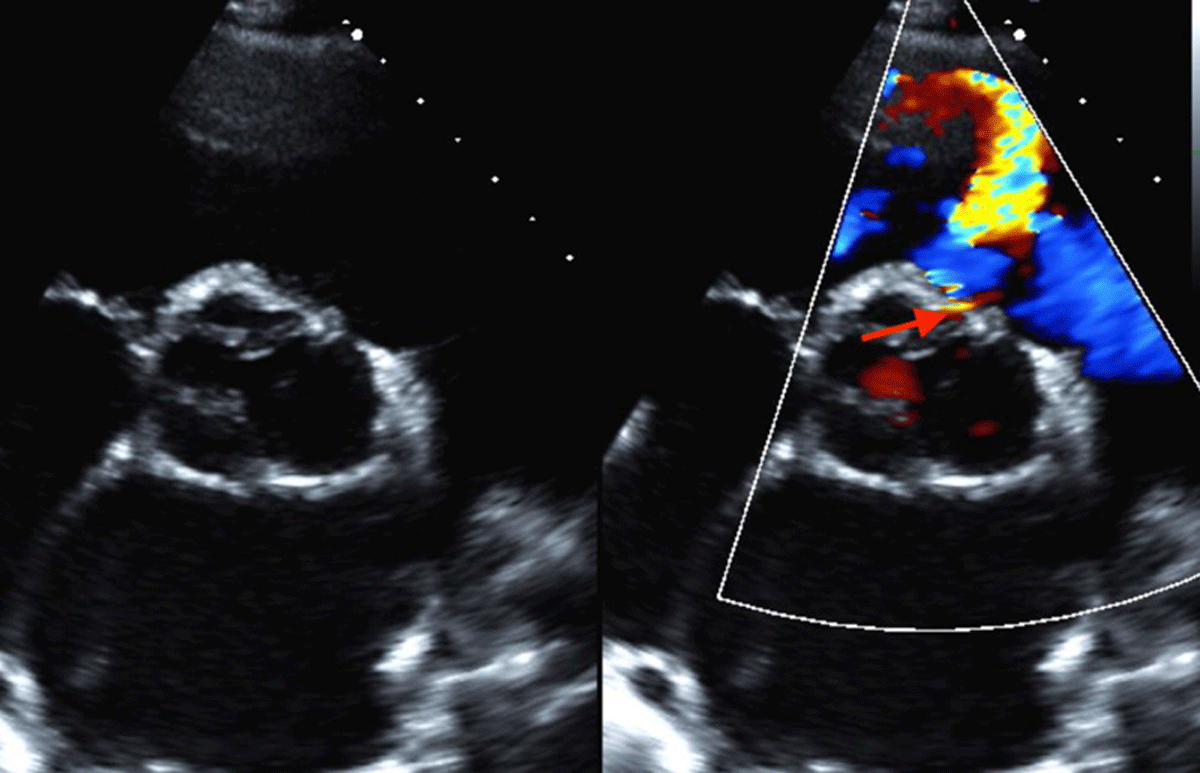
Figure 1
Transesophageal echocardiography showing ventricular septal defect jet between the 12:00 and 2:00 positions (red arrow).
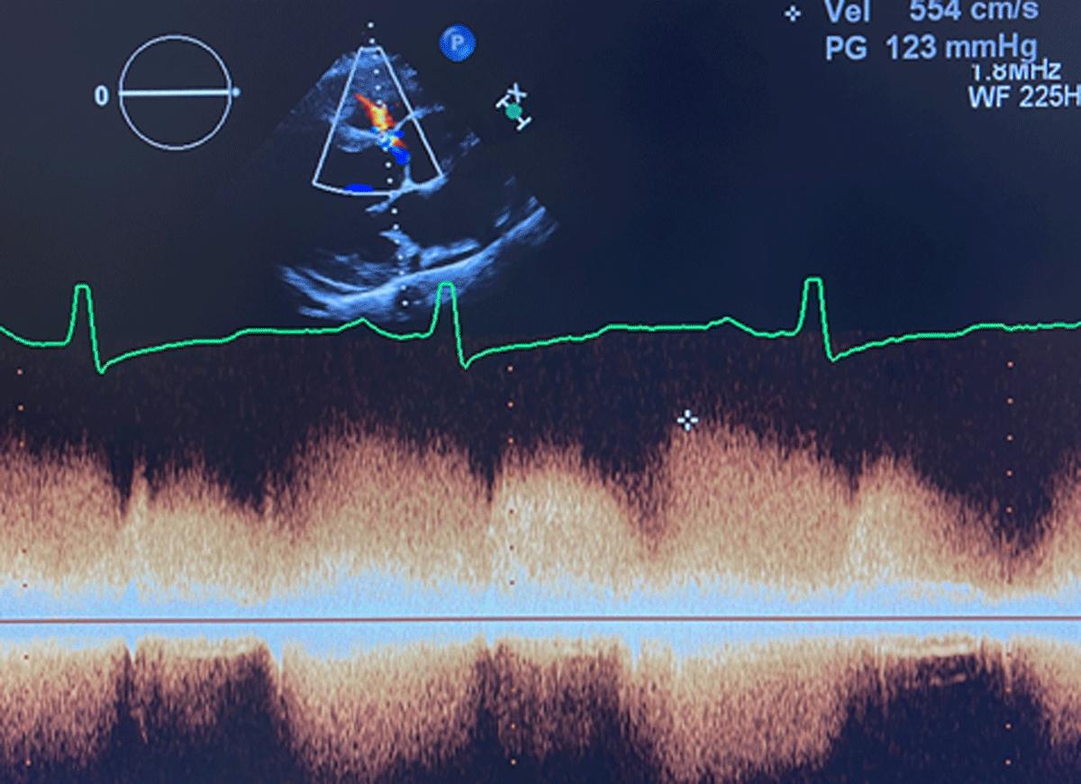
Figure 2
Doppler wave form on transesophageal echocardiogram.

Figure 3
Severe biventricular dilation seen on cardiac magnetic resonance imaging.
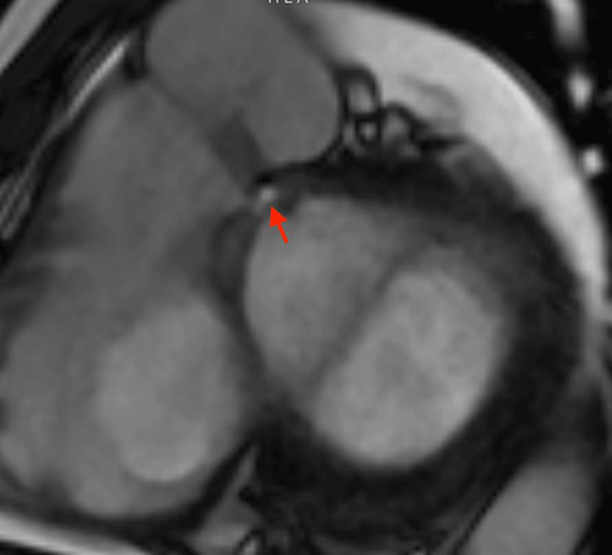
Figure 4
Supracristal ventricular septal defect (red arrow) with an 8.5-mm defect adjacent to the right sinus of Valsalva.
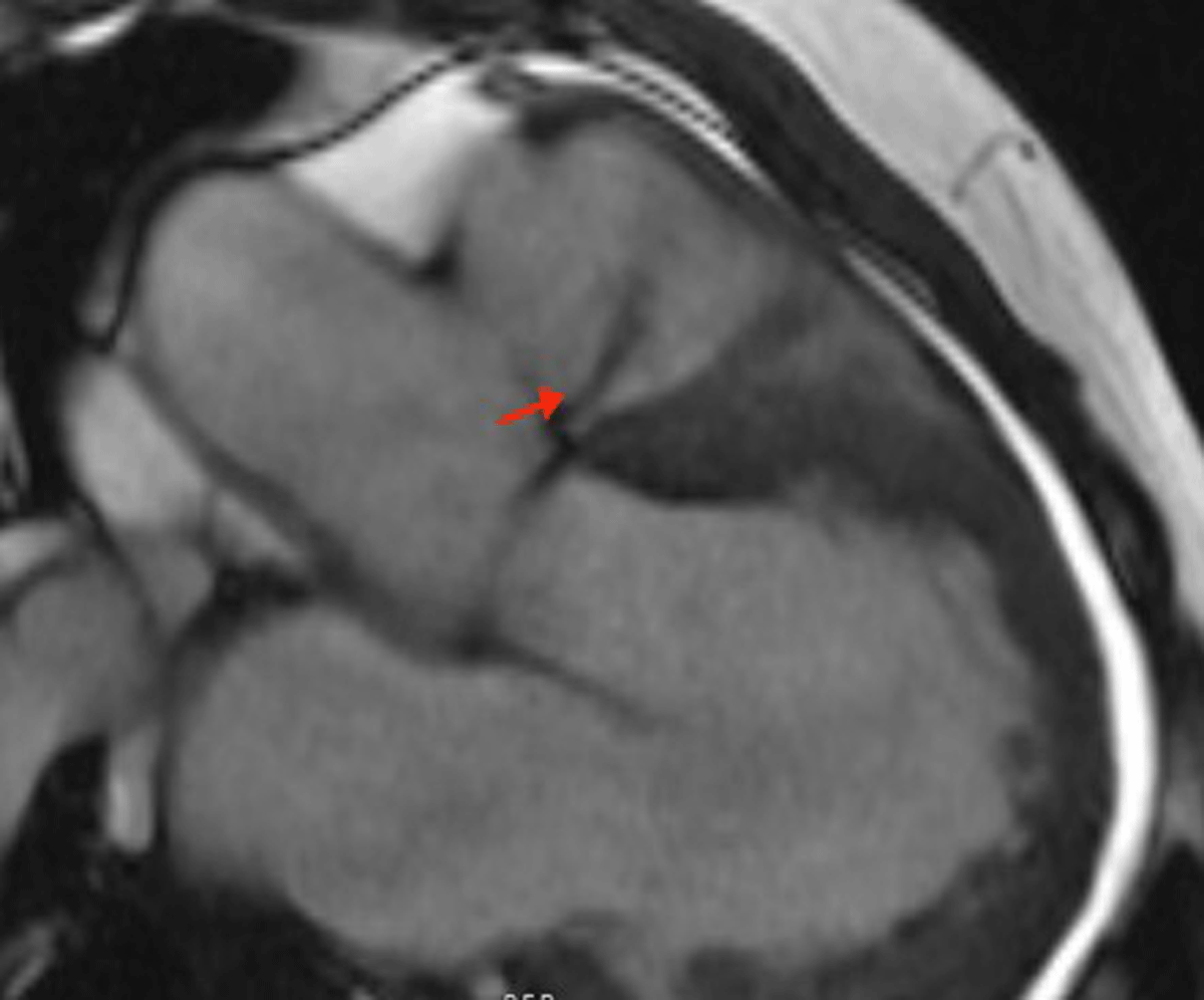
Figure 5
Aorta-right ventricular outflow tract fistula demonstrated by the presence of systolic flow jet (red arrow).

Figure 6
Aorta-right ventricular outflow tract fistula demonstrated by the presence of diastolic flow jet (red arrow).
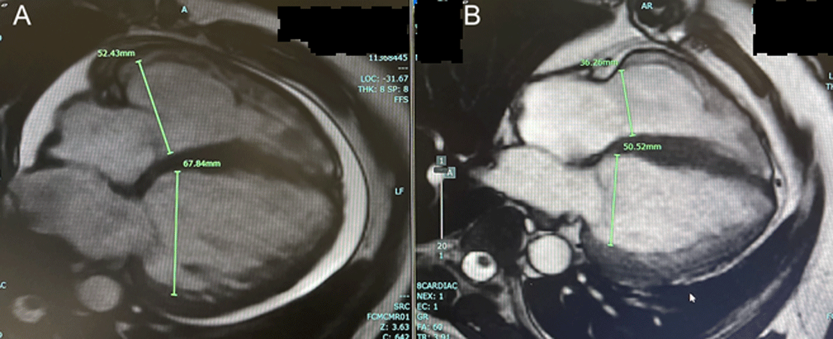
Figure 7
(A) Biventricular dilation prior to surgery. (B) After surgery, there was marked improvement in end diastolic diameters of both the left and right ventricles.