
Table 1
Updated clinical classification of pulmonary hypertension. Reproduced with permission of ©ERS 2020; DOI: 10.1183/13993003.01913-2018.3 PAH: pulmonary arterial hypertension; HIV: human immunodeficiency virus; CCB: calcium channel blockers; PVOD: pulmonary veno-occlusive disease; PCH: pulmonary capillary hemangiomatosis; LVEF: left ventricular ejection fraction; PH: pulmonary hypertension
| GROUP 1: PAH | GROUP 2: PH DUE TO LEFT HEART DISEASE |
|---|---|
|
|
| GROUP 3: PH DUE TO LUNG DISEASE AND/OR HYPOXIA | GROUP 4: PH DUE TO PULMONARY ARTERY OBSTRUCTIONS |
|
|
| GROUP 5: PH WITH UNCLEAR AND/OR MULTIFACTORIAL MECHANISMS | |
|
Table 2
Updated classification of drugs and toxins associated with pulmonary hypertension. Reproduced with permission of the ©ERS 2020: DOI: 10.1183/13993003.01913-2018.3 HCV: hepatitis C virus
| DEFINITE | POSSIBLE |
|---|---|
| Aminorex Fenfluramine Dexfenfluramine Benfluorex Methamphetamines Dasatinib Toxic rapeseed oil | Cocaine Phenylpropanolamine L-tryptophan St John’s wort Amphetamines Interferon alpha and beta Alkylating agents Bosutinib Direct-acting anti-HCV agents Leflunomide Idarubicin (Chinese herb Qing-Dai) |
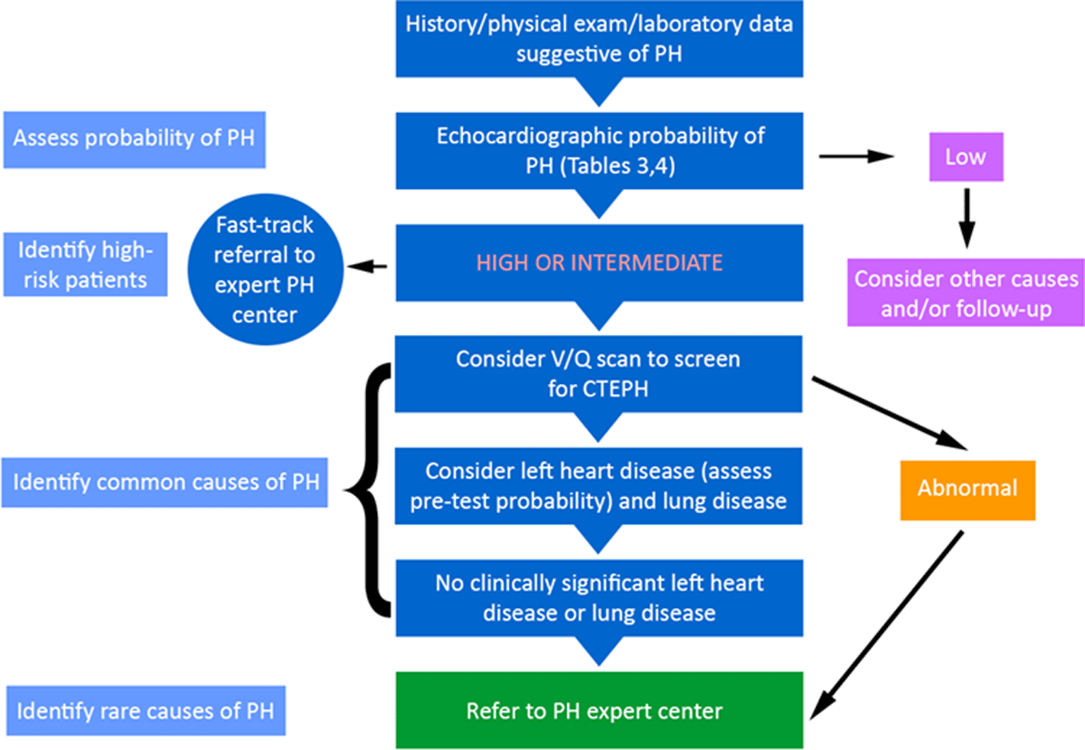
Figure 1
Algorithm for diagnosis of pulmonary hypertension and identifying its cause(s). Reproduced with permission of the © ERS 2020: DOI: 10.1183/13993003.01904-2018.7 PH: pulmonary hypertension; V/Q: ventilation/perfusion; CTEPH: chronic thromboembolic PH
Table 3
Echocardiographic probability of pulmonary hypertension (PH) in symptomatic patients with a suspicion for PH. Reproduced with permission of the ©ERS 2020: DOI: 10.1183/13993003.01904-2018.7
| PEAK TRICUSPID REGURGITANT VELOCITY M/S | PRESENCE OF OTHER ECHOCARDIOGRAPHIC “PH SIGNS” | ECHOCARDIOGRAPHIC PROBABILITY OF PH |
|---|---|---|
| ≤ 2.8 or not measurable | No | Low |
| ≤ 2.8 or not measurable | Yes | Intermediate |
| 2.9–3.4 | No | |
| 2.9–3.4 | Yes | High |
| > 3.4 | Not required |
Table 4
Echocardiographic signs that are used with tricuspid regurgitant velocity measurements in Table 3 to assess the probability of pulmonary hypertension (PH). Reproduced with permission of the ©ERS 2020: DOI: 10.1183/13993003.01904-2018.7 Echocardiographic signs from at least two different categories (A/B/C) should be present to alter the level of echocardiographic probability of PH. RA: right atrium; RV: right ventricle; LV: left ventricle; PA: pulmonary artery.
| A: THE VENTRICLES | B: PULMONARY ARTERY | C: INFERIOR VENA CAVA AND RA |
|---|---|---|
| RV/LV basal diameter ratio >
1 Flattening of the interventricular septum (LV eccentric index > 1.1 in systole and/or diastole) | RV outflow Doppler acceleration time
< 105 ms and/or mid-systolic
notching Early diastolic pulmonary regurgitation velocity > 2.2 m/s PA diameter > 25 mm | Inferior vena cava diameter > 21 mm
with decreased inspiratory collapse (< 50% with a sniff or
< 20% with quiet inspiration) RA area (end systole) > 18 cm2 |