
INTRODUCTION
Pulmonary hypertension (PH) refers to elevated pressures in the pulmonary circulation that are caused by either pulmonary vascular remodeling and inflammation or increased downstream pressures (precapillary vs postcapillary PH). The World Symposium on Pulmonary Hypertension (WSPH) meets every 5 years to update the classification, diagnosis, and management of pulmonary hypertension.1 The most recent WSPH, held in 2018, made several changes, one of which was modifying the clinical classification of PH into five groups.
There is a growing appreciation for the role that PH plays in confounding and complicating various cardiac, pulmonary, and systemic diseases. Pulmonary arterial hypertension (PAH), classified as group 1 PH, is a chronic, progressive condition that leads to elevated pressures in the pulmonary vasculature. This is characterized by abnormal smooth muscle proliferation and endothelial dysfunction, which leads to increased pulmonary vascular resistance (PVR) and eventual right heart failure. Early recognition and accurate diagnosis are of paramount importance to guide timely and appropriate management of these patients. In this review, we focus on the most updated classification, diagnosis, and evaluation of PH.
NEW CLINICAL CLASSIFICATION OF PH
The World Symposium on PH has a classification system for PH to accurately communicate about the different PH entities, standardize the diagnostic workup and treatment of PH, and conduct trials on homogenous patient groups.2 The current classification recognizes five groups of PH (Table 1).3 Group 1 is pulmonary arterial hypertension (PAH), a rare condition with a prevalence of around 15 to 50 patients per million in Europe and the United States.2 PAH can be heritable, idiopathic, drug/toxin-induced, or linked to a variety of conditions, including portal hypertension, connective tissue diseases, congenital heart disease, and human immunodeficiency virus (HIV) infection.3 There are two subgroups within group 1 that have significant differences. The first subgroup includes patients who have positive vasoreactivity testing and a long-term sustained response to calcium channel blocker treatment. It is important to identify these patients because they have a different therapeutic pathway and tend to have a better prognosis compared to other group 1 patients.3,4 The second subgroup is patients with pulmonary veno-occlusive disease (PVOD) and pulmonary capillary hemangiomatosis (PCH); these patients have a significantly poorer prognosis and are prone to developing life-threatening pulmonary edema upon initiation of PAH-specific medications.3,5 A list of drugs/toxins that have a definite or possible association with PAH is found in Table 2.
Table 1
Updated clinical classification of pulmonary hypertension. Reproduced with permission of ©ERS 2020; DOI: 10.1183/13993003.01913-2018.3 PAH: pulmonary arterial hypertension; HIV: human immunodeficiency virus; CCB: calcium channel blockers; PVOD: pulmonary veno-occlusive disease; PCH: pulmonary capillary hemangiomatosis; LVEF: left ventricular ejection fraction; PH: pulmonary hypertension
| GROUP 1: PAH | GROUP 2: PH DUE TO LEFT HEART DISEASE |
|---|---|
|
|
| GROUP 3: PH DUE TO LUNG DISEASE AND/OR HYPOXIA | GROUP 4: PH DUE TO PULMONARY ARTERY OBSTRUCTIONS |
|
|
| GROUP 5: PH WITH UNCLEAR AND/OR MULTIFACTORIAL MECHANISMS | |
|
Table 2
Updated classification of drugs and toxins associated with pulmonary hypertension. Reproduced with permission of the ©ERS 2020: DOI: 10.1183/13993003.01913-2018.3 HCV: hepatitis C virus
| DEFINITE | POSSIBLE |
|---|---|
| Aminorex Fenfluramine Dexfenfluramine Benfluorex Methamphetamines Dasatinib Toxic rapeseed oil | Cocaine Phenylpropanolamine L-tryptophan St John’s wort Amphetamines Interferon alpha and beta Alkylating agents Bosutinib Direct-acting anti-HCV agents Leflunomide Idarubicin (Chinese herb Qing-Dai) |
Group 2 PH includes patients who have left heart disease, group 3 includes those with chronic lung diseases and/or hypoxia, and group 4 patients have pulmonary artery obstructions, including chronic thromboembolic PH (CTEPH). Group 5 is composed of a heterogenous group with unknown or multifactorial mechanisms of PH development (Table 1).1,3
EVALUATION AND DIAGNOSIS OF PH
There is a well-documented time lag between symptom onset and diagnosis of PH. According to the Registry to Evaluate Early and Long-Term PAH Disease Management (REVEAL registry), roughly 21% of patients experienced a 2-year delay in receiving a diagnosis of PAH.6 The lack of improvement in this lag over the past decades triggered the 6th WSPH taskforce on the diagnosis of PH to modify the recommended diagnostic algorithm to facilitate a more efficient diagnostic process.7 For example, patients with PH due to confounding medical conditions are typically medically complex and difficult to treat, prompting the recommendation that they receive early referral to specialized PH centers to guide their long-term management (Figure 1).6,8,9,10 In the United States, these centers are called Pulmonary Hypertension Care Centers and are accredited by the Pulmonary Hypertension Association.11 Care received at PH care centers has been shown to improve outcomes in patients with PAH.12
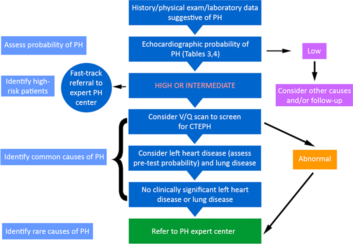
Figure 1
Algorithm for diagnosis of pulmonary hypertension and identifying its cause(s). Reproduced with permission of the © ERS 2020: DOI: 10.1183/13993003.01904-2018.7 PH: pulmonary hypertension; V/Q: ventilation/perfusion; CTEPH: chronic thromboembolic PH
History and Physical Examination Findings
All patients suspected to have PH undergo a thorough history taking and physical examination with an emphasis on findings that may point to the presence of PH or associated conditions. PH usually presents with nonspecific symptoms of exertional dyspnea, chest pain, generalized weakness and fatigue, light-headedness or syncope in advanced cases, and symptoms of right ventricular (RV) failure and systemic fluid overload.7,13 Physical exam findings suggestive of PH include prominent P2, right ventricular heave, jugular venous distension, hepatojugular reflux, peripheral edema, tricuspid or pulmonary regurgitation murmurs, and S3 gallop in the absence of crackles and wheezing on pulmonary auscultation.7,13
Diagnostic Tests
Electrocardiography
Patients suspected of having PH undergo a comprehensive workup to confirm the presence of PH and identify its etiology. One of the initial tests obtained from these patients is electrocardiography (EKG). Although a normal EKG does not rule out PH, EKG changes in PH are nonspecific and include right axis deviation, right bundle branch block, RV hypertrophy, P pulmonale, and QTc prolongation.14 The EKG findings associated with a poor prognosis include increased P wave amplitude in lead II, P ≥ .25 mV in lead II, qR in V1, and the presence of World Health Organization criteria for RV hypertrophy.14,15
Laboratory tests
Blood tests are usually obtained during initial evaluation for PH. Abnormalities may indicate conditions associated with or causative of PH or signal end-organ compromise, a consequence of PH. Thyroid disease is common in PAH and should be checked along with routine hematological and biochemical testing. Screening for connective tissue diseases, hepatitis, and HIV is required.7 Recommended serological testing for scleroderma includes ANA immunofluorescence (considered positive if ≥ 1:160). When there is high clinical suspicion, the physician should consider a panel that includes anticentromere, antitopoisomerase, anti-RNA polymerase III, double-stranded DNA, anti-Ro, anti-La, and U1-RNP antibodies.7 Elevated brain natriuretic peptide (BNP) or N-terminal pro-BNP indicate RV overload and predict poor outcomes. In cases where a thrombophilic state is suspected, as in CTEPH and connective tissue diseases, physicians should perform coagulopathy and thrombophilia screenings, including anticardiolipin antibodies, lupus anticoagulant, and β2-glycoprotein antibodies.7
Pulmonary function tests
In patients with PAH, the most common abnormalities in pulmonary function testing are reduced diffusing capacity of the lung for carbon monoxide (DLCO) and mild restrictive pattern. The presence of overt obstructive or restrictive patterns indicates the presence of underlying airway or lung parenchymal diseases.16, 17, 18 Significant reductions in DLCO to < 60% predicted or the presence of marked exertional hypoxemia warrant consideration of PVOD/PCH.19 Cardiopulmonary exercise testing is also used for evaluation and follow-up of PH patients because it allows one to quantify the degree of relative hypoperfusion of the lungs and systemic circulation during exercise, grade the severity of exercise limitation, and assess the response to therapy.20,21
Ventilation/perfusion scans
Ventilation/perfusion (V/Q) scans have the greatest screening utility in evaluation of CTEPH. It is necessary to obtain a V/Q scan in all patients undergoing evaluation for PH. A normal V/Q scan rules out CTEPH, which can be a cause of unexplained dyspnea.22 Thus, patients presenting with unexplained dyspnea must undergo V/Q testing.
Chest computed tomography
Chest computed tomography (CT) can provide several clues to the presence and etiology of PH. Findings of dilated right atrium (RA) or RV or main pulmonary artery (PA) are suggestive of PH. The presence of interstitial lung disease may suggest lung disease as the etiology for PH, and the magnitude of parenchymal involvement is taken into consideration to discriminate between group 1 vs group 3 PH.7
Transthoracic echocardiography
This is the most important noninvasive screening test for PH, and suggestive findings are then confirmed by invasive testing with right heart catheterization (RHC).7 The echocardiographic probability of PH is based on tricuspid regurgitant velocity, RV size, interventricular septal function, inferior vena cava diameter respiratory variation, systolic RA area, pattern of systolic flow velocity and early diastolic pulmonary regurgitant velocity, and diameter of the PA (Tables 3, 4).23, 24, 25, 26 In addition, an echo saline bubble study should be performed to exclude right-to-left shunting.
Table 3
Echocardiographic probability of pulmonary hypertension (PH) in symptomatic patients with a suspicion for PH. Reproduced with permission of the ©ERS 2020: DOI: 10.1183/13993003.01904-2018.7
| PEAK TRICUSPID REGURGITANT VELOCITY M/S | PRESENCE OF OTHER ECHOCARDIOGRAPHIC “PH SIGNS” | ECHOCARDIOGRAPHIC PROBABILITY OF PH |
|---|---|---|
| ≤ 2.8 or not measurable | No | Low |
| ≤ 2.8 or not measurable | Yes | Intermediate |
| 2.9–3.4 | No | |
| 2.9–3.4 | Yes | High |
| > 3.4 | Not required |
Table 4
Echocardiographic signs that are used with tricuspid regurgitant velocity measurements in Table 3 to assess the probability of pulmonary hypertension (PH). Reproduced with permission of the ©ERS 2020: DOI: 10.1183/13993003.01904-2018.7 Echocardiographic signs from at least two different categories (A/B/C) should be present to alter the level of echocardiographic probability of PH. RA: right atrium; RV: right ventricle; LV: left ventricle; PA: pulmonary artery.
| A: THE VENTRICLES | B: PULMONARY ARTERY | C: INFERIOR VENA CAVA AND RA |
|---|---|---|
| RV/LV basal diameter ratio >
1 Flattening of the interventricular septum (LV eccentric index > 1.1 in systole and/or diastole) | RV outflow Doppler acceleration time
< 105 ms and/or mid-systolic
notching Early diastolic pulmonary regurgitation velocity > 2.2 m/s PA diameter > 25 mm | Inferior vena cava diameter > 21 mm
with decreased inspiratory collapse (< 50% with a sniff or
< 20% with quiet inspiration) RA area (end systole) > 18 cm2 |
Right heart catheterization
PH is defined hemodynamically, and the 6th WSPH changed the definition of PH to mean pulmonary artery pressure (mPAP) ≥ 20 mm Hg. Precapillary PH (group I PAH) is defined by mPAP ≥ 20 mm Hg, pulmonary capillary wedge pressure (PCWP) ≤ 15 mm Hg, and pulmonary vascular resistance (PVR) ≥ 3 Wood units (WU) on an RHC. Although there is evidence that even PVR > 2 WU is considered abnormal in all age groups,27 the current hemodynamic definition of PAH recognizes a cutoff of PVR ≥ 3 WU, which is somewhat arbitrarily defined.3,28 All patients suspected to have PH must undergo RHC, which is associated with relatively low morbidity and mortality (1.1% and 0.055%, respectively) when performed at experienced centers.29 All pressure measurements are taken at end-expiration of tidal breathing. Pressures are recorded at the RA, RV, PA, and PA wedge.
It can be difficult to obtain accurate wedge pressure tracing in patients with severe PAH. In these cases, wedge position should be confirmed with saturation (which should proximate left atrial saturation). In rare cases, left ventricular end diastolic pressure measurement should be considered with arterial puncture. Cardiac output (CO) is measured by the thermodilution method or the direct Fick method; thermodilution is considered the gold standard given the inaccuracies with the Fick method, which is commonly used.30 Oxygen saturation is measured at least in the superior vena cava, RA, and PA; if greater than 75% in the PA, it should be measured in each cardiac chamber to assess for left-to-right shunting.23 Other derived parameters are used to differentiate precapillary from postcapillary PH and include the pulmonary vascular resistance (PVR = [mPAP-PAWP]/CO), transpulmonary gradient (TPG = mPAP-PAWP), and diastolic pressure gradient (DPG = mean PAWP-diastolic PAP). Vasoreactivity testing is recommended only for patients with idiopathic, heritable, and drug-induced PAH.3,31 The test is performed using inhaled nitric oxide at 10 ppm to 20 ppm as the preferred agent, but intravenous epoprostenol or adenosine or inhaled iloprost may be used as alternatives. Acute response is defined as a reduction in mPAP of at least 10 mm Hg to reach an mPAP of 40 mm Hg or less, with unchanged or increased cardiac output.3
Genetic Testing
It is important to note that several genetic mutations have been identified that increase the risk of developing PAH.32 These mutations have been identified in idiopathic PAH, anorexigen-induced PAH, and in idiopathic and familial PVOD/PCH. Patients diagnosed with any of these should be informed of the possibility that other family members may have an increased risk of developing the same disorder and given the option of screening asymptomatic family members.
Genetic counseling is an essential component of managing patients with PAH. The 6th WSPH specifies that patients should be offered genetic counseling before undergoing genetic testing to discuss concepts of inheritance patterns and varied penetrance. Asymptomatic family members who choose to undergo screening should be tested for the mutation identified in their affected family member; testing negative for that mutation means their risk of developing PAH is similar to that of the general population.32
NOVEL IMAGING MODALITIES
There is accumulating evidence for the utility of incorporating other imaging modalities to evaluate for PH. These include dual energy CT for assessment of pulmonary perfusion,33 V/Q single photon emission CT for improved accuracy at excluding pulmonary embolism,34,35 magnetic resonance imaging (MRI) techniques to assess lung perfusion and ventilation, and cardiac MRI-based RV strain imaging.36,37 The availability and widespread use of these modalities at specialized PH centers remains variable. An updated PH diagnostic algorithm is outlined in Figure 1.7
SCREENING OF HIGH-RISK POPULATIONS
Several patient populations are at an increased risk of developing PH, including patients with connective tissue diseases, HIV infection, congenital heart disease, portopulmonary hypertension, and hereditary hemorrhagic telangiectasia.6 Accordingly, screening for PH is recommended in these groups to allow timely diagnosis and treatment. Asymptomatic carriers of mutations associated with PAH should be screened with an annual echocardiogram.
CONCLUSION
Despite current knowledge of its pathophysiology and the development of more therapeutic options, PH is a complex disease that carries a high risk of morbidity and mortality and requires detailed evaluation and management at an expert center. The most recent WSPH revised the clinical classification of PH into five groups. Screening for PH is recommended for high-risk groups, such as patients with HIV infection and congenital heart disease. Other novel modalities are being incorporated into the workup of PH patients, and this is an evolving field. Early referral to a specialized PH center is strongly recommended.
KEY POINTS
Comprehensive evaluation for pulmonary arterial hypertension (PAH) must be done at an expert pulmonary hypertension (PH) center.
Right heart catheterization is essential for diagnosis of PH.
A newer hemodynamic definition must be considered for early diagnosis and treatment of PAH.
In patients with hereditary PAH, family genetic screening is encouraged.
Conflict of Interest Disclosure
Dr. Beshay reports a research grant from ACCP CHEST. Dr. Sahay reports personal fees and nonfinancial support from Bayer Pharmaceuticals, United Therapeutics, Actelion Pharmaceuticals, personal fees from Liquidia, Gossamer Bio, and Altavant sciences, and a grant from ACCP CHEST. He is also a member of the clinical trial end point adjudication committee of a randomized clinical trial sponsored by GlaxoSmithKline.