
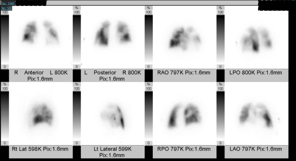
Figure 1
Ventilation-perfusion scan of patient with thromboembolic pulmonary hypertension showing multiple wedge-shaped perfusion deficits bilaterally.
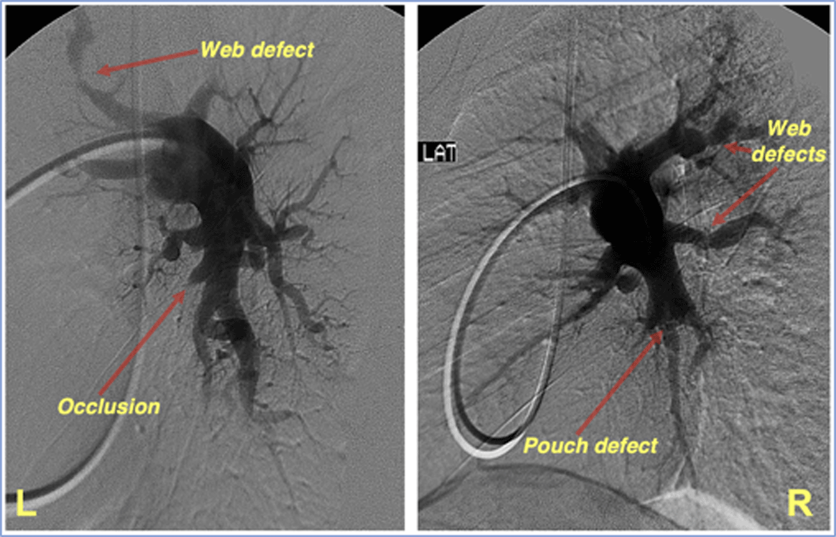
Figure 2
Digital subtraction angiograms demonstrating multiple webs, vessel occlusions, and pouch defects consistent with thromboembolic pulmonary hypertension.

Figure 3
(A) Coronal and (B) axial computed tomography demonstrating a web-like filling defect in a 52-year-old female with thromboembolic pulmonary hypertension.
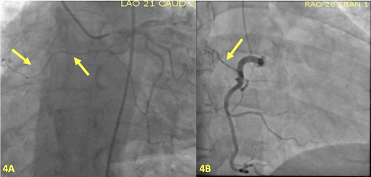
Figure 4
Bronchial collateralization from (A) the left circumflex artery and from (B) the conus branch of the right coronary artery.
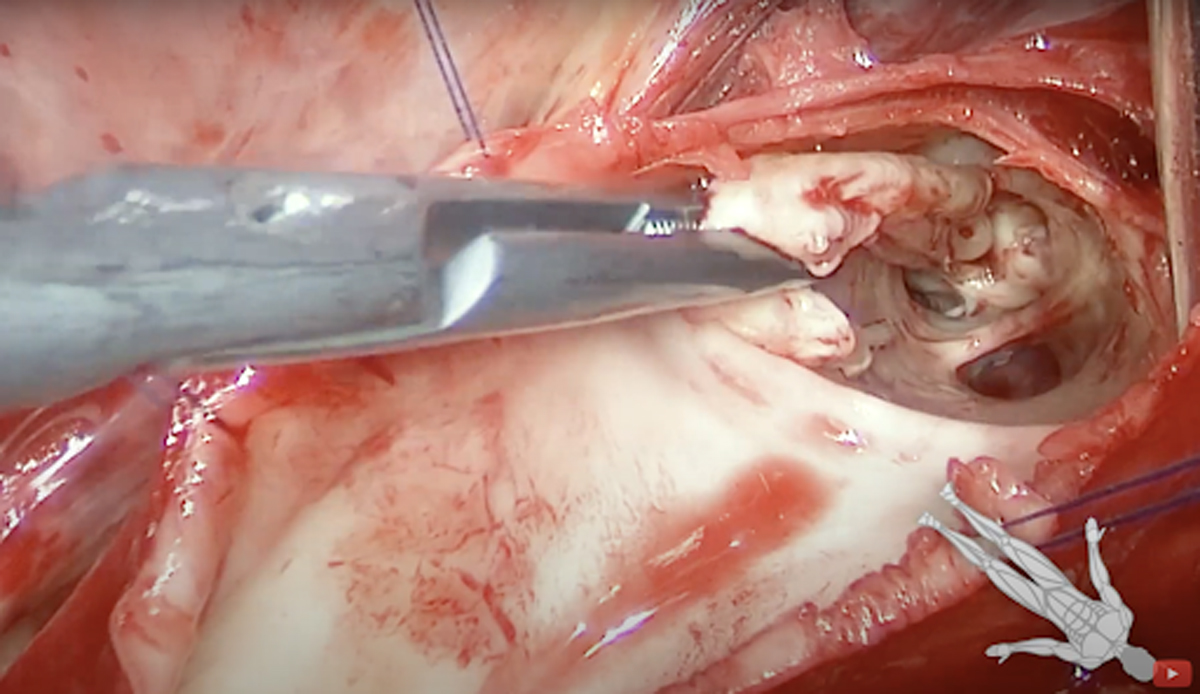
Figure 5
View from right pulmonary vasculature demonstrating the importance of a bloodless field to achieve complete distal dissection in pulmonary thromboendarterectomy.
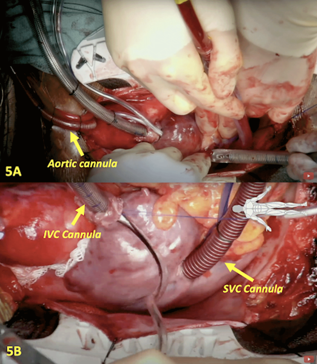
Figure 6
(A) Central aortic cannulation for cardiopulmonary bypass. (B) Recommended configuration for bicaval venous cannulation (IVC/SVC) to facilitate mobilization of the SVC and exposure of the RPA medially. IVC: inferior vena cava; SVC: superior vena cava; RPA: right pulmonary artery.
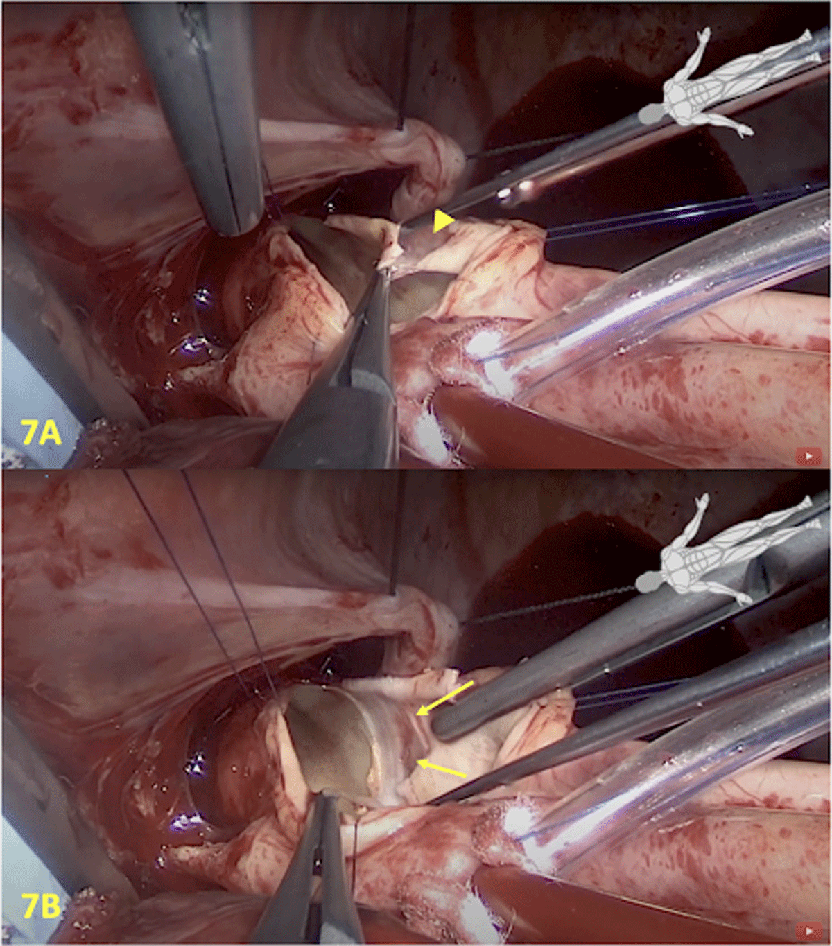
Figure 7
(A) The correct dissection plane is white and smooth and often offers little resistance. (B) Yellow arrows indicate an incorrect plane of dissection, which appears pink.
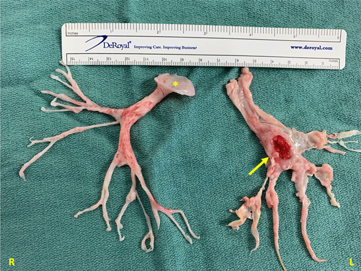
Figure 8
Specimen from a 37-year-old male with a history significant for antiphospholipid syndrome presenting with complete occlusion of right pulmonary artery, which appears mushroom-like (asterisk). Left pulmonary endarterectomy specimen shows an ulcerated lesion (arrow).