
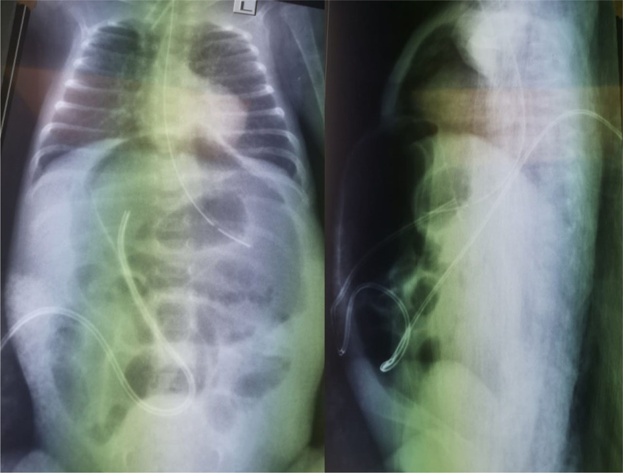
Fig. 1.
Abdominal X-ray: (A) Anterior-posterior view: thickened, dilated bowel loops marked with arrow (B) Lateral decubitus view: pneumoperitoneum – marked with arrow
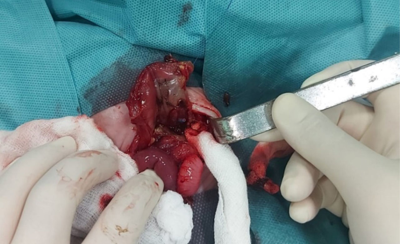
Fig. 2.
Intraoperative finding: gastric wall perforation with the presence of the nasogastric tube tip (circled)