
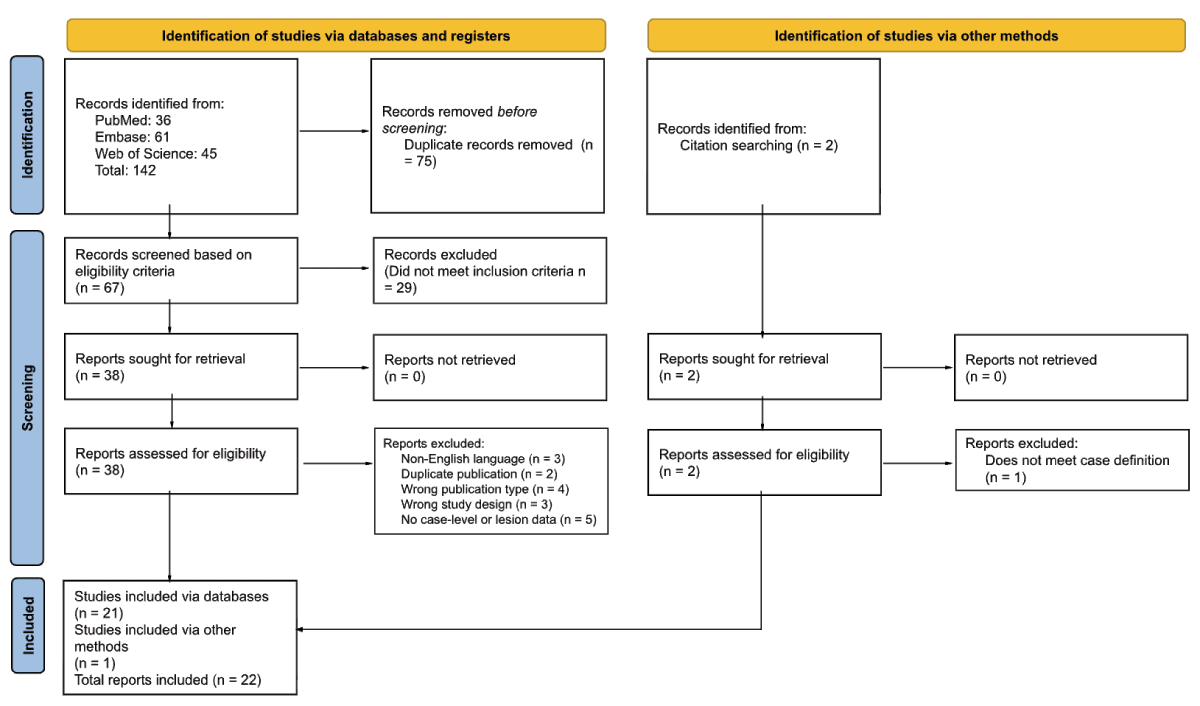
Figure 1
PRISMA Flowchart. Study identification, screening, and inclusion process, adapted from Page MJ, et al. BMJ 2021;372:n71. doi: 10.1136/bmj.n71.
Table 1
Clinical and neuroanatomical features of reported parakinesia brachialis oscitans cases. Cases are ordered chronologically by year of publication. Full case-level data are given in Supplementary Table 3. CADASIL, cerebral autosomal dominant arteriopathy with subcortical infarcts and leukoencephalopathy; ICA, internal carotid artery; MCA, middle cerebral artery; NR, not reported; PBO, parakinesia brachialis oscitans; PCA, posterior cerebral artery.
| # | AUTHOR (YEAR) | AGE/SEX | LESION/DIAGNOSIS | SYMPTOM LATERALITY | LIMB(S) AFFECTED | TIME TO ONSET | PBO OUTCOME |
|---|---|---|---|---|---|---|---|
| 1 | Blin et al. (1994) | 62/M | Left posterior limb of internal capsule infarct | Right | Upper & Lower | A few days | Persisted at 6 weeks |
| 2 | Töpper et al. (2003) | 62/M | Right PCA and MCA territory infarction | Left | Upper | NR | NR |
| 3 | Töpper et al. (2003) | 43/M | Basilar artery thrombosis with right pontine infarct | Left | Upper | 4 days | NR |
| 4 | Töpper et al. (2003) | 51/M | Hypertensive left thalamic hemorrhage extending into the internal capsule | Right | Upper | 2 weeks | NR |
| 5 | Walusinski et al. (2010) | 49/F | Right internal capsule infarct in the setting of CADASIL (NOTCH3 mutation) | Left | Upper | 2 days | Resolved at 2 weeks with motor recovery |
| 6 | Walusinski et al. (2010) | 73/M | Left internal capsule lacunar infarct | Right | Upper | At presentation | Resolved at 1 week with partial recovery and residual ataxia |
| 7 | Walusinski et al. (2010) | 71/M | Right centrum semiovale and lenticular nucleus infarct | Left | Upper | At presentation | Persisted at 3 years |
| 8 | Walusinski et al. (2010) | 53/M | Right caudate and centrum semiovale infarct in the setting of factor V Leiden | Left | Upper | At presentation | Persisted at 1 year |
| 9 | Walusinski et al. (2010) | 75/M | Left internal capsule and lenticular nucleus infarct | Right | Upper | At presentation | NR |
| 10 | Walusinski et al. (2010) | 35/F | Left total MCA territory infarct in the setting of ICA dissection and fibromuscular dysplasia | Right | Upper | At presentation | NR |
| 11 | de Lima et al. (2012) | 63/M | Right cortical and subcortical frontoparietal infarct | Left | Upper | 2 months | NR |
| 12 | Jung et al. (2012) | 59/M | Right precentral gyrus and frontal subcortical infarct | Left | Upper | 1 day | Resolved, timing NR, by 5-year follow-up |
| 13 | Zorzetto et al. (2013) | 60/M | Left MCA territory infarct | Right | Upper | During thrombolysis | Resolved at 24 hours |
| 14 | Wu et al. (2013) | 52/M | Right putaminal hemorrhage extending into the internal capsule | Left | Upper | 4 months | Persisted, increasing in frequency and intensity |
| 15 | Farah et al. (2015) | 51/M | Left fronto-parieto-temporal infarct | Right | Upper | 2 days | Resolved at 12 hours |
| 16 | Kang and Dhand (2015) | 63/M | Left cortical and subcortical infarct in the MCA territory | Right | Upper | NR | Resolved at 2 weeks with partial recovery of arm strength |
| 17 | Koo and Wang (2016) | 57/F | Right internal capsule infarct | Left | Upper | A few days | NR |
| 18 | Nascimento Alves et al. (2018) | 59/M | Left lenticular nucleus, internal capsule, caudate and corona radiata infarct | Right | Upper | At presentation | NR |
| 19 | Aaron and Al Hashmi (2019) | 53/F | Right pontine and upper medullary infarct | Left | Upper | 2 days | Resolved at 3 days |
| 20 | Gadelha et al. (2019) | 65/F | Right MCA territory infarct with M1 occlusion | Left | Upper | During thrombolysis | NR |
| 21 | Chowdhury et al. (2022) | 59/M | Left frontal cortex and basal ganglia infarct with hemorrhagic transformation | Right | Upper | 2 weeks | Persisted at 1 year with reduced amplitude |
| 22 | Lim and Neo (2022) | 65/M | Right corona radiata and internal capsule infarct | Left | Upper | 17.5 hours | Resolved, timing NR, with recovery of motor power |
| 23 | Salavisa et al. (2023) | 35/M | Right centrum semiovale demyelinating lesion; multiple sclerosis | Left | Upper | 2 weeks | Improved, then re-emerged with neurological worsening |
| 24 | Zobiri et al. (2023) | 71/F | Internal capsule and lenticular nucleus infarct, right MCA territory | Left | Upper | NR | NR |
| 25 | Tsai et al. (2024) | 31/M | Right deep subcortical white matter tumefactive demyelinating lesion | Left | Upper | At presentation | NR |
| 26 | Alvarez and Alvarez (2025) | 76/M | Right frontal cortical-subcortical intracerebral hemorrhage | Left | Upper | NR | NR |
| 27 | Orozco-Puga et al. (2026) | 20/F | Right internal capsule infarct in the setting of ICA dissection | Left | Upper | 1 week | NR |
| 28 | Orozco-Puga et al. (2026) | 45/F | Left pontine infarct with hemorrhagic transformation | Right | Upper | 4 days | NR |
| 29 | Orozco-Puga et al. (2026) | 49/F | Right frontal subcortical white matter infarct | Left | Upper | 2 weeks | NR |
| 30 | AlJaafari and Shariff (2026) | 57/M | Left pontine infarct with basilar atherosclerosis | Right | Upper & Lower | 1 week | Resolved by 3 months |
| 31 | Alcalá-Torres (2026) | 76/F | Right lenticular nucleus and internal capsule infarct | Left | Upper | 1 month | Resolved at 5 months |
Table 2
Summary of clinical and lesion characteristics in parakinesia brachialis oscitans. Values are n (%) unless otherwise stated. Percentages are calculated on the denominator stated for each block. Associated neurological features and anatomical levels are not mutually exclusive. Onset beyond one month ranged from 2 to 4 months. Among cases resolving after more than one week, time to resolution ranged from 2 weeks to 5 months. Among cases that persisted, follow-up extended to 3 years.
| VARIABLE | N (%) |
|---|---|
| Sex (n = 31) | |
| Men | 21 (67.7) |
| Women | 10 (32.3) |
| Age at onset, years (n = 31) | |
| Mean (SD) | 56.1 (13.6) |
| Range | 20 to 76 |
| Laterality of symptoms (n = 31) | |
| Left | 19 (61.3) |
| Right | 12 (38.7) |
| Limb involvement (n = 31) | |
| Upper only | 29 (93.5) |
| Upper and lower | 2 (6.5) |
| Associated neurological features (n = 31) | |
| Dysarthria | 10 (32.3) |
| Facial weakness | 10 (32.3) |
| Aphasia | 6 (19.4) |
| Sensory abnormalities | 3 (9.7) |
| Headache | 3 (9.7) |
| Limb incoordination | 2 (6.5) |
| Reduced level of consciousness | 2 (6.5) |
| Vertigo | 1 (3.2) |
| Visual field deficit | 1 (3.2) |
| Seizures | 1 (3.2) |
| Time to PBO onset (n = 31) | |
| At presentation | 9 (29.0) |
| 1 to 7 days | 11 (35.5) |
| More than 1 week to 1 month | 5 (16.1) |
| More than 1 month | 2 (6.5) |
| Not specified | 4 (12.9) |
| Anatomical level involved (n = 27) | |
| Internal capsule | 12 (44.4) |
| Corona radiata or subcortical white matter | 11 (40.7) |
| Basal ganglia | 8 (29.6) |
| Cortex | 6 (22.2) |
| Brainstem | 4 (14.8) |
| Thalamus | 1 (3.7) |
| Etiology (n = 31) | |
| Acute ischemic stroke | 26 (83.9) |
| Intracerebral hemorrhage | 3 (9.7) |
| Inflammatory or demyelinating lesion | 2 (6.5) |
| Outcome (n = 31) | |
| Resolved within 1 week | 4 (12.9) |
| Resolved after more than 1 week | 4 (12.9) |
| Resolved, timing not reported | 2 (6.5) |
| Improved but not resolved | 1 (3.2) |
| Persisted at last follow-up | 5 (16.1) |
| Not reported | 15 (48.4) |
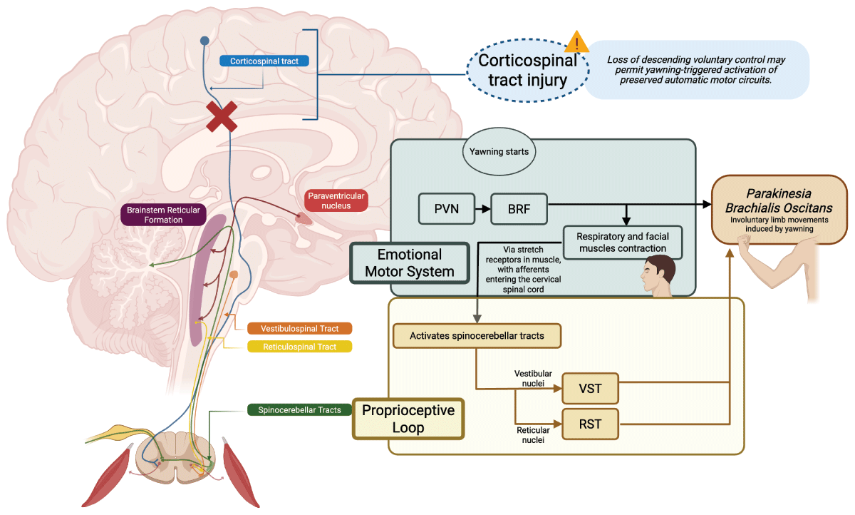
Figure 2
Proposed mechanistic framework for parakinesia brachialis oscitans. In a limb already weakened by a focal lesion interrupting descending voluntary control, yawning is accompanied by involuntary movement that may reflect activation of preserved automatic motor circuits. Two hypothesized pathways are illustrated. Neither has been tested directly in patients with PBO. In the emotional motor system pathway, yawning is accompanied by activation of the paraventricular nucleus (PVN) and the brainstem reticular formation (BRF), with contraction of respiratory and facial muscles, and involuntary limb movements via intact reticulospinal projections. In the proprioceptive loop pathway, muscle stretch receptors in respiratory and limb muscle and tendon send afferent signals into the cervical spinal cord and ascend in the spinocerebellar tracts, which in turn engage the vestibular and reticular nuclei, with descending motor output through the vestibulospinal tract (VST) and reticulospinal tract (RST). Both pathways are proposed routes to the involuntary yawning-triggered limb movements characteristic of PBO. BRF, brainstem reticular formation; PVN, paraventricular nucleus; RST, reticulospinal tract; VST, vestibulospinal tract.