
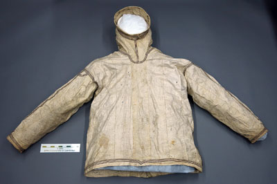
Figure 1
Parka N: 233 before conservation treatment. It was collected in Tasiilaq in 1930–1, and was donated to the museum in 1939. Photo: ©Scott Polar Research Institute, University of Cambridge.
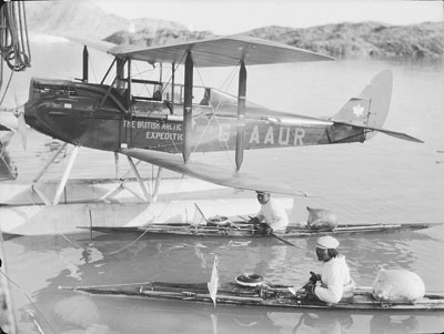
Figure 2
Inuit kayakers with an aircraft from the British Arctic Air Route expedition (BAARE) 1930–1. Photo: ©Scott Polar Research Institute, University of Cambridge.
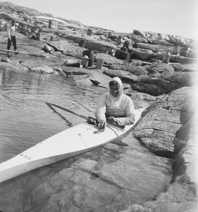
Figure 3
A member of the BAARE expedition (possibly Henry Iliffe Cozens) wearing a gut parka while kayaking in Greenland 1930–1. Photo: ©Scott Polar Research Institute, University of Cambridge.
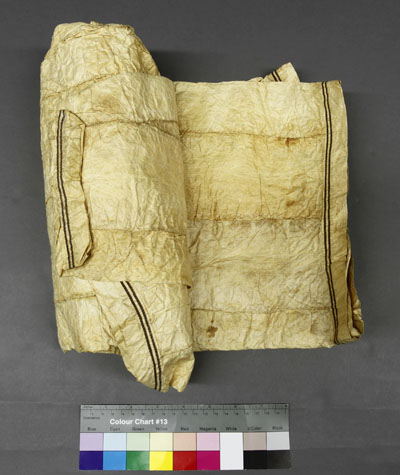
Figure 4
The Wager parka was rolled up tightly when it first arrived at the museum in 2011. Photo: ©Scott Polar Research Institute, University of Cambridge.
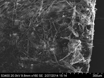
Figure 5
SEM image of the inner surface of the gut from the Wager parka at 160× magnification. Micrograph: University College London.
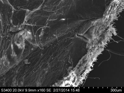
Figure 6
SEM image of the outer surface of the gut from the Wager parka at 160× magnification. Micrograph: University College London.
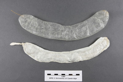
Figure 7
Comparing sausage skin which has been air dried (top) as opposed to freeze dried (bottom). Freeze drying resulted in a more opaque and silvery appearance. Photo: ©Scott Polar Research Institute, University of Cambridge.
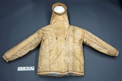
Figure 8
The Wager parka unfolded after humidification and reshaping. Photo: ©Scott Polar Research Institute, University of Cambridge.
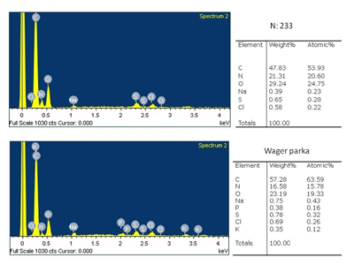
Figure 9
Results of SEM EDX elemental analysis for both parkas. No iron was detected in either one. Spectrograph: University College London.
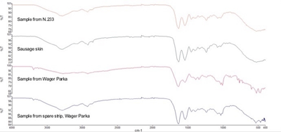
Figure 10
Results of FTIR spectroscopy. Spectrograph: University College London.
Table 1
Table of FTIR wavelength peaks (cm–1). Results are shown from the two parkas, the spare strip from the Wager parka, and a sausage skin reference sample.
| Sausage skin reference sample | Sample from N: 233 | Sample from Wager Parka | Sample from spare strip, Wager Parka |
|---|---|---|---|
| 3694 ± 2 | 3692 ± 2 | ||
| 3293 | 3284 ± 2 | 3280 ± 1 | 3281 ± 1 |
| 3202 ± 18 | |||
| 3086 | 3075 ± 3 | 3072 ± 12 | 3073 ± 1 |
| 2922 | 2924 ± 1 | 2924 ± 3 | 2926 ± 4 |
| 2854 | 2852 ± 3 | 2861 ± 10 | 2850 ± 0 |
| 1629 | 1633 ± 1 | 1634 ± 2 | 1633 ± 3 |
| 1540 | 1538 ± 1 | 1538 ± 2 | 1539 ± 4 |
| 1457 | 1451 ± 0 | 1442 ± 5 | 1450 ± 1 |
| 1402 | 1401 ± 1 | 1389 ± 3 | 1391 ± 1 |
| 1336 | 1335 ± 2 | 1340 ± 3 | 1335 ± 2 |
| 1236 | 1235 ± 1 | 1238 ± 3 | 1234 ± 1 |
| 1084 | 1079 ± 1 | 1082 ± 1 | 1081 ± 2 |
| 1035 | 1032 ± 2 | 1031 ± 0 | 1030 ± 2 |
| 1007 ± 0 | |||
| 884 ± 3 | |||
| 722 ± 0 |
Table 2
Table comparing the relative intensity (AI/AII) and relative locations (Δv) of the Amide I and II peaks, corresponding to the degree of hydrolyzation and oxidation of the collagen structure. Results are shown for the two parkas and the sausage skin reference sample.
| Sample | AI/AII | Δv |
|---|---|---|
| Sausage skin reference | 89 | 0.98 |
| N: 233 | 95 | 0.93 |
| Wager Parka | 96 | 0.93 |
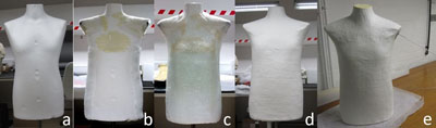
Figure 11
The process of building up the lightweight paper torso. A: The basic polystyrene mannequin used as a former. B: Plastazote and polyester wadding added to pad out the former to the desired shape and size. C: Cling film wrapped over the whole surface to form a separation layer. D: The paper shell being built up over the mannequin former. E: The paper mount after removal from the former with one metal armature attached through the shoulder. Photo: ©Scott Polar Research Institute, University of Cambridge.
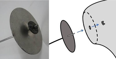
Figure 12
Structure of the sleeve armature showing the metal rod with fixed large plate and wing nut to attach the rod through the shoulder of the paper torso. Photo: ©Scott Polar Research Institute, University of Cambridge.
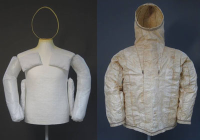
Figure 13
The completed mount (left), and the Wager parka on the mount (right). Photo: ©Scott Polar Research Institute, University of Cambridge.