
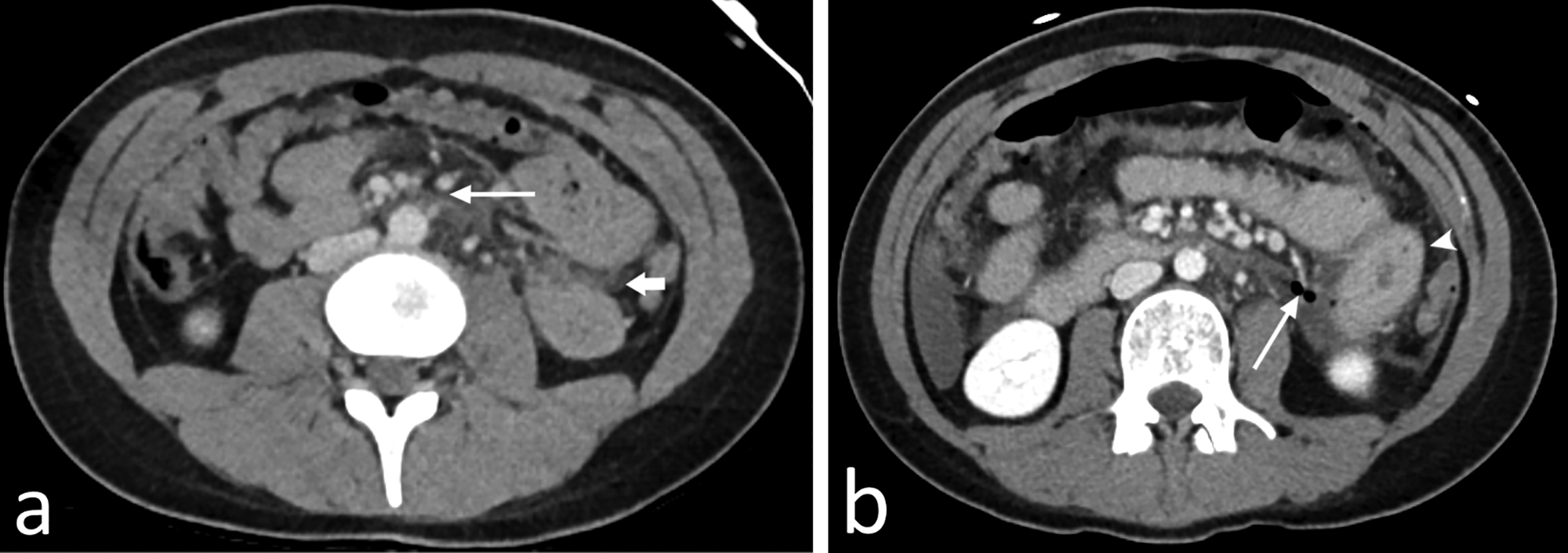
Figure 1
Axial (a) CT in the venous phase of an 18‑year‑old woman who was involved in a car accident at very high speed (200 km/h). She underwent whole‑body CT scan, which only showed subtle mesenteric infiltration central in the abdomen (long arrow) and a minimal amount of free fluid on the left side (short arrow) and in the pouch of Douglas (not shown). Because she was stable and alert and had no abdominal pain, she was admitted for observation. Later, she developed severe abdominal pain, for which a repeat CT scan (b) was performed approximately 10 hours after the initial scan. There is an increased amount of free fluid, a massive pneumoperitoneum with several small air bubbles posteriorly in the abdomen (long arrow), and an adjacent small bowel loop with significant wall thickening (arrowhead). Exploratory laparoscopy confirmed jejunal perforation.