
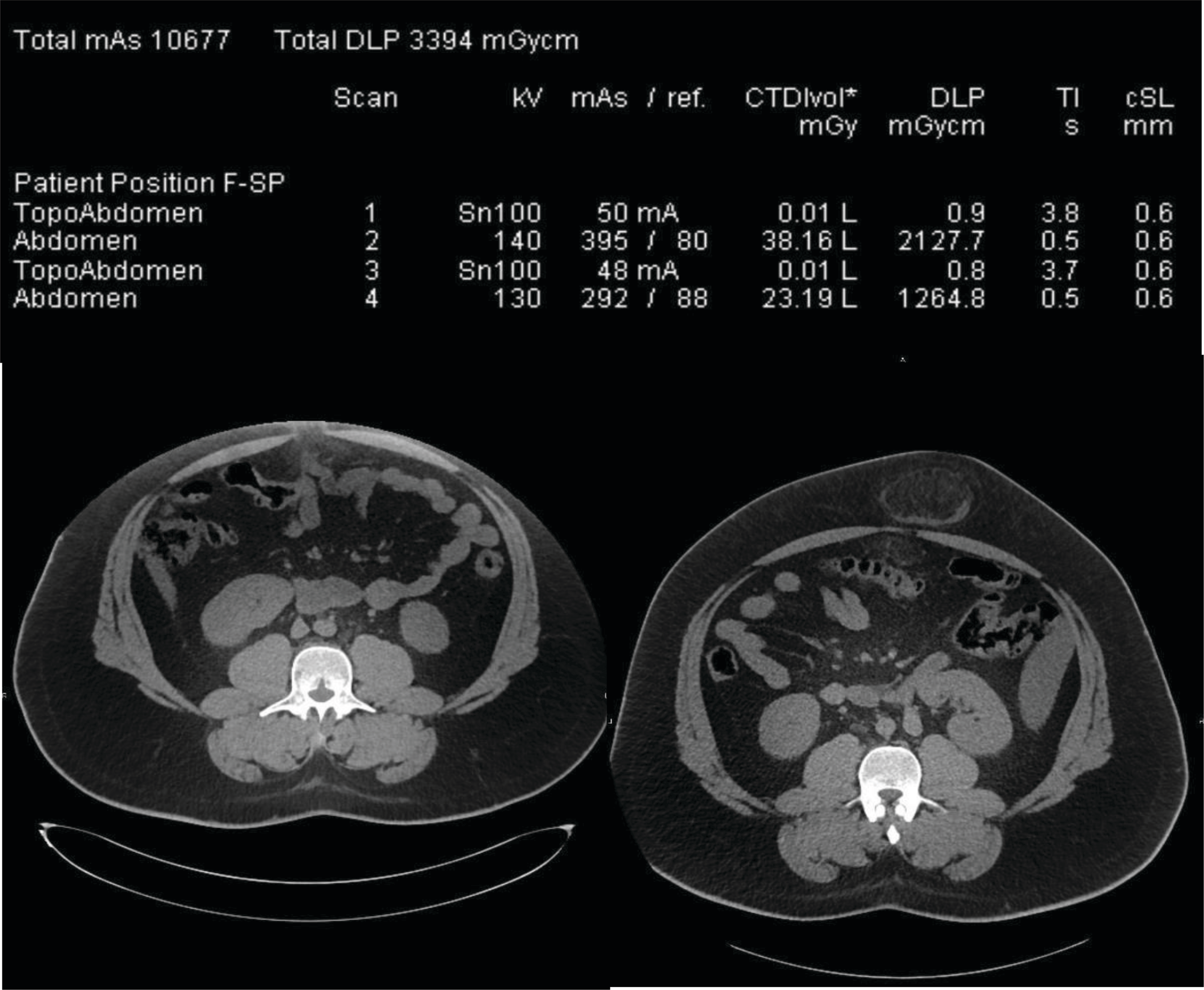
Figure 1
Radiation dose report and CT slices obtained at the level of the umbilic in a 24 year‑old patient. The left CT slice is off‑centered and the herniation is not visible whereby the right CT slice of correctly centered and the herniation is visible. The two first exposures on the dose report correspond to the left image whereby the two last lines correspond to the right image including the umbilical herniation.