
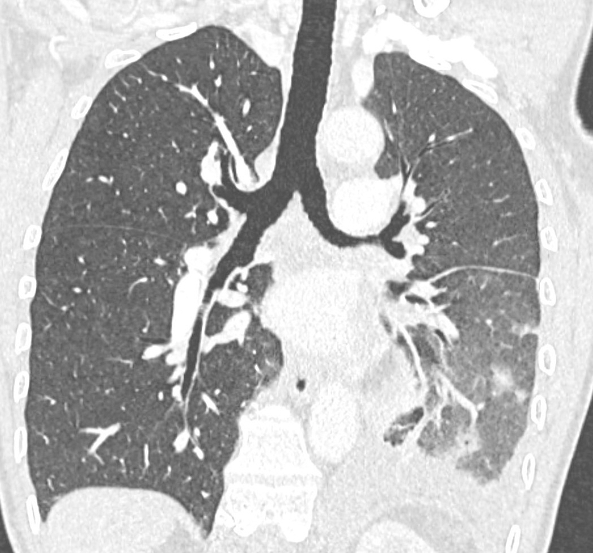
Figure 1
Contrast‑enhanced coronal chest CT (pulmonary window) with unilateral left signs of patchy alveolar hemorrhages, pulmonary edema, and pleural effusion.
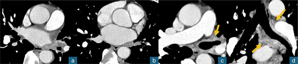
Figure 2
Contrast‑enhanced axial chest CT (mediastinal window); a, b: thrombosis of the left superior and inferior pulmonary veins; c, d: diffuse left peribronchial contrast‑enhancing infiltration (arrows), “pulmonary hilar cavernoma.”
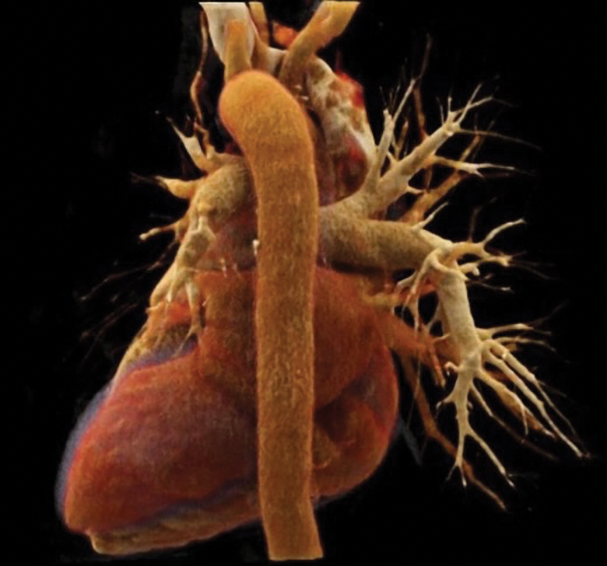
Figure 3
Cinematic VRT posterior view of the mediastinum; neither upper nor lower left pulmonary veins (in red color) are patent.