

Figure 1 A‑B.
Conventional radiography shows an elongated, well circumscribed heterotopic ossification projecting superolaterally to the anterior inferior iliac spine (AIIS) (arrows). There is also cam deformity with loss of concavity of the femoral head‑neck junction.
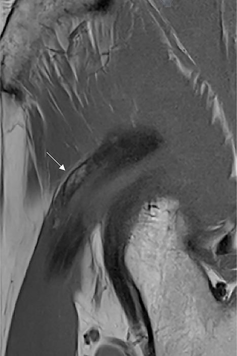
Figure 2
Magnetic Resonance Imaging shows mature HO containing fatty bone marrow along the course of the indirect head of the rectus femoris (arrow) on sagittal T1‑weighted images.
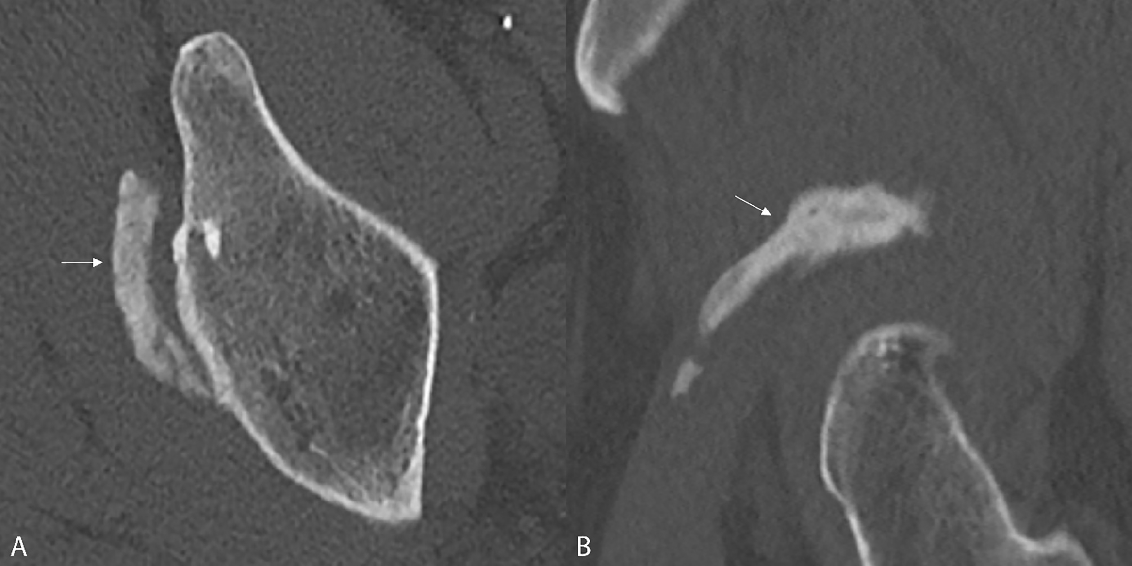
Figure 3 A‑B.
Computed Tomography confirms HO with elongated morphology superior to the acetabular rim (arrows).