Have a personal or library account? Click to login
Hereditary Leiomyomatosis and Renal Cell Cancer (HLRCC) Syndrome Cover

Hereditary Leiomyomatosis and Renal Cell Cancer (HLRCC) Syndrome

Open Access
|Sep 2024

Figures & Tables

jbsr-108-1-3687-g1.jpg
Figure 1

Expansile mass in the upper portion of the left kidney (thick arrows in A, B, D), with invasion of the sinusal fat tissue (B, D). A delayed nephrogram is apparent secondary to outflow obstruction due to renal vein thrombosis (thin arrow in C). Large, confluent, retroperitoneal lymphadenopathies can be seen (arrowheads in B, C).

jbsr-108-1-3687-g2.jpg
Figure 2

Gross specimen of the resected kidney.
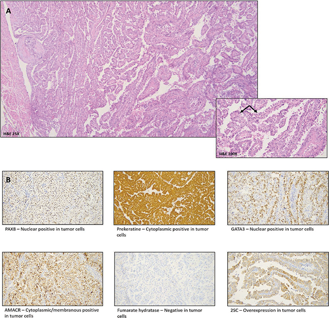
jbsr-108-1-3687-g3.jpg
Figure 3

A. H&E – Encapsulated tumor with tubulopapillary architecture. Tumor cells show eosinophilic cytoplasm and pleomorphic hyperchromatic nuclei with Fuhrmann nuclear grade 3. B. Immunohistochemistry markers with PAX8/panCK positivity.
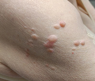
jbsr-108-1-3687-g4.jpg
Figure 4

Multiple skin papules/nodules on the upper arm, compatible with cutaneous leiomyoma.

DOI: https://doi.org/10.5334/jbsr.3687 | Journal eISSN: 2514-8281
Language: English
Submitted on: Jul 3, 2024
Accepted on: Aug 22, 2024
Published on: Sep 11, 2024
Published by: Ubiquity Press
In partnership with: Paradigm Publishing Services
Publication frequency: 1 issue per year

© 2024 Karel Mercken, Brecht Van Berkel, Liesbeth De Wever, published by Ubiquity Press
This work is licensed under the Creative Commons Attribution 4.0 License.