
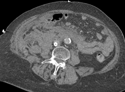
Figure 1
An axial CT of extensive intra- and retroperitoneal blood at the level of the infrarenal aorta.
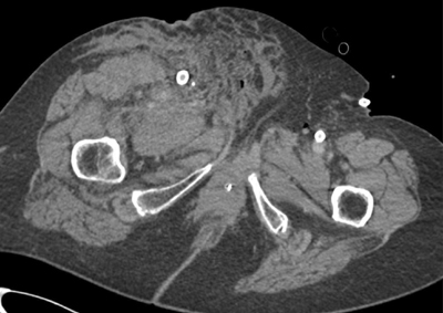
Figure 2
An axial CT showing extensive inguinal blood at the level of the right superficial femoral artery.

Figure 3
Paracoronal reformatted image of the watershed zone (white arrows) at the level of the superior mesenteric artery (SMA).
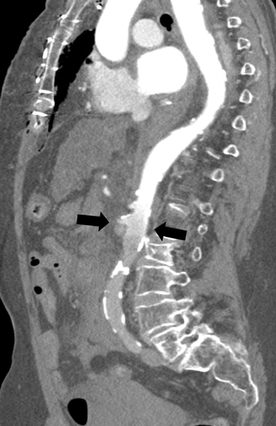
Figure 4
Parasagittal reformatted image of the watershed zone (black arrows) at the level of the superior mesenteric artery (SMA).

Figure 5
3D reconstruction of the watershed zone at the level of the superior mesenteric artery (SMA).
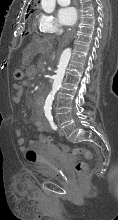
Figure 6
Parasagittal image of the contrast-enhanced aorta.
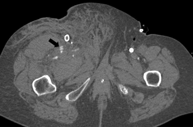
Figure 7
An axial image of a clear arterial contrast blush from the superficial femoral artery and a side branch.