
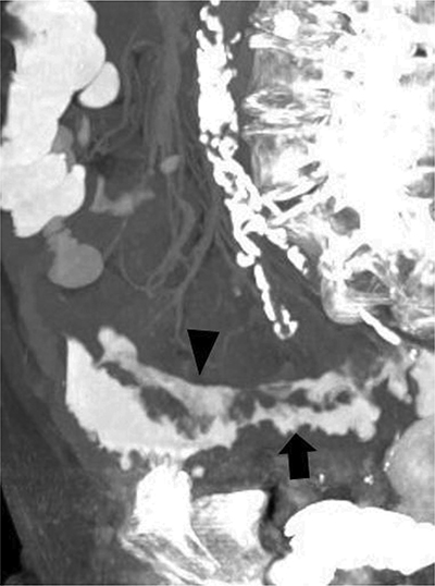
Figure 1
Maximum intensity projection imaging of the abdominal CT: two contrast trajectories are distinguishable, the intramural longitudinal fistula (arrowhead) and the normal intraluminal contrast of the sigmoid (arrow).
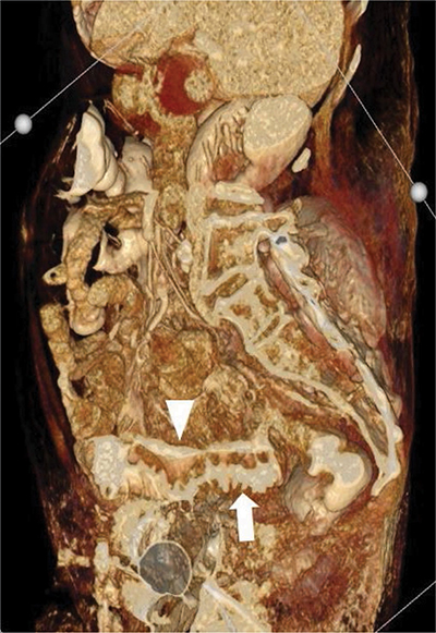
Figure 2
Volume-rendered three-dimensional reconstruction of the abdominal CT. The intramural longitudinal fistula is shown (arrowhead) to run parallel with the intraluminal contrast of the sigmoid (arrow).