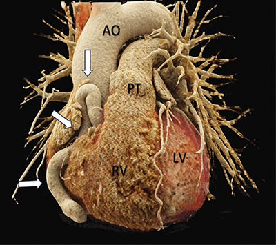
Have a personal or library account? Click to login
Right Coronary Artery-to-Coronary Sinus Fistula with Giant Tortuous Dilatation of the Right Coronary Artery Cover

Right Coronary Artery-to-Coronary Sinus Fistula with Giant Tortuous Dilatation of the Right Coronary Artery

Open Access
|Jun 2024

Figures & Tables

jbsr-108-1-3546-g1.jpg
Figure 1

3D rendered cardiac CT-angiography reveals a tortuous dilation of the right coronary artery (white arrows). Front view.

Source: AO = Aorta, PT = Pulmonary Trunk, RV = Right ventricle, LV = Left Ventricle.
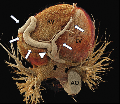
jbsr-108-1-3546-g2.jpg
Figure 2

3D rendered cardiac CT-angiography reveals a tortuous dilation of the right coronary artery (white arrows). Near the origin of the posterior interventricular artery, a coronary artery fistula is visible (white arrowhead) that communicates with a moderately dilated coronary sinus (black arrows). Bottom view.

jbsr-108-1-3546-g3.jpg
Figure 3

Cardiac CT-angiography reveals a coronary artery-to-coronary sinus fistula (white arrowhead). Note the dilation of the right coronary artery (white arrow). Axial view.

DOI: https://doi.org/10.5334/jbsr.3546 | Journal eISSN: 2514-8281
Language: English
Submitted on: Feb 12, 2024
Accepted on: May 14, 2024
Published on: Jun 3, 2024
Published by: Ubiquity Press
In partnership with: Paradigm Publishing Services
Publication frequency: 1 issue per year

© 2024 Louis Waterloos, Charles Edouard Heylen, published by Ubiquity Press
This work is licensed under the Creative Commons Attribution 4.0 License.