
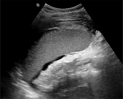
Figure 1
Abdominal ultrasound shows splenomegaly and a perisplenic, layered fluid collection.
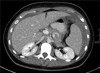
Figure 2
Contrast-enhanced abdominal CT shows a haemoperitoneum. A hyperdense, perisplenic collection with layering suggests an acute perisplenic haematoma.
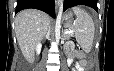
Figure 3
Contrast-enhanced abdominal CT shows a haemoperitoneum and splenomegaly. A hyperdense, perisplenic suggests an acute perisplenic haematoma.